
An official website of the United States government
The .gov means it’s official. Federal government websites often end in .gov or .mil. Before sharing sensitive information, make sure you’re on a federal government site.
The site is secure. The https:// ensures that you are connecting to the official website and that any information you provide is encrypted and transmitted securely.
- Publications
- Account settings
Preview improvements coming to the PMC website in October 2024. Learn More or Try it out now .
- Advanced Search
- Journal List
- Int J Mol Sci

The Etiology of Cholelithiasis in Children and Adolescents—A Literature Review
Katarzyna zdanowicz.
1 Department of Pediatrics, Gastroenterology, Hepatology, Nutrition and Allergology, Medical University of Bialystok, 15-274 Bialystok, Poland
Jaroslaw Daniluk
2 Department of Gastroenterology and Internal Medicine, Medical University of Bialystok, 15-276 Bialystok, Poland
Dariusz Marek Lebensztejn
Urszula daniluk, associated data.
Not applicable.
The incidence of gallstone disease has increased in recent years. The pathogenesis of cholelithiasis is not fully understood. The occurrence of the disease is influenced by both genetic and environmental factors. This article reviews the literature on cholelithiasis in children, with the exception of articles on hematological causes of cholelithiasis and cholelithiasis surgery. The aim of this review is to present the latest research on the pathogenesis of gallstone disease in children. The paper discusses the influence of all factors known so far, such as genetic predisposition, age, infections, medications used, parenteral nutrition, and comorbidities, on the development of gallstone disease. The course of cholelithiasis in the pediatric population is complex, ranging from asymptomatic to life-threatening. Understanding the course of the disease and predisposing factors can result in a faster diagnosis of the disease and administration of appropriate treatment.
1. Introduction
The broad spectrum of biliary tract disease includes the most often diagnosed gallstone disease (cholelithiasis), cholecystitis, or biliary dyskinesia [ 1 , 2 ]. Gallstones are formed mainly in the gallbladder, less often in intrahepatic or extrahepatic bile ducts. More patients with cholelithiasis have no obvious symptoms. Symptomatic patients presented with dyspepsia and biliary colic caused mainly by obstruction of the cystic duct [ 3 ]. Gallstones may lead to serious complications such as cholecystitis, acute cholangitis, and pancreatitis [ 1 ]. In recent years, the prevalence of cholelithiasis has risen and ranges from 1.9% to 4% in children [ 4 , 5 ]. This increase may be caused by the worrying problem of childhood obesity and the widespread use of ultrasound [ 6 , 7 , 8 ]. Simultaneously, in pediatric patients, the number of performed cholecystectomies rose by 213% over a 9-year period [ 9 ]. Additionally, cases of fetal cholelithiasis were also described [ 10 , 11 ]. Schwab et al., in approximately 200,000 obstetric sonograms taken between 1996 and 2019, found 34 fetal cholelithiases. The median gestational age at diagnosis was 34.7 weeks, and the majority of cases were female (56%) [ 10 ]. Troyano-Luque et al. reported two cases of fetal cholelithiasis. In one of them, cholelithiasis was diagnosed 6 weeks after 16 days of ceftriaxone therapy in maternal Lyme disease [ 11 ]. The presence of gallstones in fetuses may lead researchers to look for genetic causes of the disease.
Based on their composition, gallstones are classified into pigment stones, cholesterol stones, and mixed stones. Pigment stones are mainly observed in hemolytic diseases, and their incidence remains stable [ 9 ]. Cholesterol gallstones are caused by genetic and environmental factors leading to an elevated concertation of cholesterol in the bile.
Cholesterol plays a structural and metabolic role. It is a component of membrane cells, steroid hormones, and a bile acid precursor. The level of cholesterol in the human body depends on endogenous de novo synthesis, enterohepatic recirculation, and dietary intake [ 12 ]. The liver is an organ that is significantly involved in cholesterol metabolism through its synthesis mediated by 3-hydroxy-3-methylglutaryl coenzyme A reductase (HMGR), cholesterol uptake by receptors (low-density lipoprotein receptors (LDLr), the prolow-density lipoprotein receptor-related protein 1 (LRP1), the scavenger receptor class B member 1 (SRB1), release of lipoproteins into the bloodstream, conversion of cholesterol into bile acids or cholesterol excretion into bile. Cholesterol is secreted by adenosine triphosphate protein binding cassette transporters G5/G8 (ABC G5/G8) to the biliary tract [ 13 ]. As a hydrophilic substance, it is solubilized in mixed micelles and vesicles that consist of bile acids and phosphatidylcholine [ 14 ].
Intestinal absorption is relevant for maintaining cholesterol balance. Cholesterol is absorbed by the Niemann–Pick C1-like 1 (NPC1L1) transporter, which, together with ABCG5 / G8, is located in the enterocyte brush border membrane. Intracellular cholesterol is esterified by the acetyl-CoA cholesterol acyltransferasen 2 (ACAT-2) and incorporated along with triglycerides, phospholipids, and apolipoprotein B-48 into chylomicrons. Next, through the lymphatic system and bloodstream, the chylomicron remnants may be absorbed by the liver. Additionally, ABCG5/G8 proteins promote the active efflux of non-esterified cholesterol and plant sterols from the enterocytes into the intestinal lumen for fecal excretion [ 15 ].
In hepatocytes, cholesterol is covered in bile acids through classical and alternative pathways. The first is the dominant one, mediated by 7α-hydroxylase (CYP21A1). The alternative pathway is controlled by sterol 27-hydroxylase (CYP27A1) and is responsible for the production of 9% to 25% of bile acids. Chenodeoxycholic acid has produced both ways, and cholic acid is only synthesized in a classic way. Newly produced bile acids, in conjunction with glycine and taurine, are pumped out by the ATP-binding cassette, sub-family B member 11 (ABC B11) [ 16 ].
Bile-binding acids are reabsorbed by the apical sodium-dependent bile acid transporter (ASBT) in the terminal ileum and then secreted into the portal vein by the organic solute transporter α/β (OST α/β). In the liver, conjugated bile acids return to the liver via Na+/taurocholic acid cotransport polypeptide (NTCP). Members of the Organic Anion Transporting Polypeptide (OATP) family are responsible for the uptake of unconjugated or sulfated bile acids. Enterohepatic circulation provides approximately 95% of bile acid return [ 16 , 17 ].
In the human body, cholesterol synthesis equals its secretion as bile acid. However, it may be disturbed by food intake and diseases [ 17 ]. The state of homeostasis is regulated by the expression of genes responsible for cholesterol transport and metabolism, transcription factors, and posttranscriptional regulatory circuits. The farnesoid X receptor (FXR) and the liver X receptors (LXRs) are nuclear receptors that play an important role in the regulation of genes encoding ABC transporters [ 18 ]. Lowering cellular cholesterol activates the endoplasmic reticulum membrane-bound transcription factor, sterol regulatory element-binding protein isoform 2 (SREBP-2), leading to enhanced cholesterol uptake and biosynthesis through regulation of genes encoding low-density lipoprotein (LDL) receptor and HMGR. On the contrary, an increase in the level of cellular cholesterol promotes (LXRs) transcription factors to enhanced cholesterol efflux from the liver [ 19 ]. The expression of CYP7A1 in the liver is regulated by the fibroblast growth factor 19 (FGF19). FGF19 is activated by bile acid in the ileum mediated by the action of ASBT. The inhibition of ASBT causes an increase in FGF19 levels and the synthesis of bile acids [ 12 ].
The risk factors for the development of cholelithiasis include age, gender, body weight, comorbidities, diet, and physical activity [ 20 ]. Although these factors are recognized in the adult population, they are modified in children. The formation of cholesterol gallstones is triggered by excessive cholesterol concentration in bile. Defects responsible for this condition are genes, hypersecretion of liver cholesterol, rapid phase transitions of cholesterol in bile, dysmotility of the gallbladder, and intestinal factors [ 20 ].
The aim of the review is to present the latest research on the pathogenesis of cholelithiasis in children. To date, many studies on the development and incidence of gallstone disease in adults have been published. To the best of our knowledge, there are no reviews of the pathomechanism of cholelithiasis development in children. A review of MEDLINE/PubMed data was carried out in August 2022, using the phrase ‘cholelithiasis’ or ‘gallstones’ in combination with ’children,’ ‘adolescents,’ or ‘pediatrics.’ Screening of the titles and abstracts was independently performed by two investigators. The studies included in this analysis fulfilled the inclusion criteria: they explored the etiology of cholelithiasis in pediatric patients and had available full texts. We excluded from the analysis articles on hematological causes of gallstone disease and surgical procedures for cholelithiasis. The selected papers were discussed with all authors.
A Swedish twin study showed that an inherited predisposition is responsible for 25% of the overall risk of developing gallstones [ 21 ]. Lithogenic genes 1 and 2 ( Lith1 and Lith2 ), playing a role in liver cholesterol secretion and regulating bile flow, have been described in murine models. Their human counterparts are ABCG5 and ABCG8 [ 17 ]. A recent study of 214 children with cholelithiasis showed the presence of the lithogenic ABCG8 allele p.D19H in 14.9% of children, which was more frequently reported compared to children and adults without gallstones. Additionally, carriers of one copy of the lithogenic variant p.D19H were also at higher risk of the development of gallstones. Increased susceptibility to the formation of cholesterol stones is associated with abnormal cholesterol metabolism resulting from its increased transport or lower intestinal absorption in combination with increased cholesterol synthesis [ 22 ]. MicroRNAs (miRNAs) are small noncoding RNAs that regulate gene expression. Recent studies have shown that miRNA-223, can prevent the development of gallstones in mice on a lithogenic diet by directly affecting the ABCG5 and ABCG8 transporters. To date, no studies have been published assessing the effect of miRNA-223 on the development of gallstone disease in humans [ 23 ].
Krawczyk et al. also found that NPC1L1 rs217434 polymorphism also was connected with the occurrence of gallstones (only in comparison with healthy adults) and lower campesterol: desmosterol ratio. However, the UGT1A1 genotype did not differ between children with and without cholelithiasis [ 22 ].
Nissinen et al. determined the D19H polymorphism of the ABCG8 gene, serum cholesterol, non-cholesterol sterols, and lipids in 66 children affected by gallstones in later life and 126 children from the control group. In the first group, 22.7% of patients carried the ABCG8 19H allele, and in the control group, this percentage was 19.0%. A decrease in phytosterols was observed in patients with the lithogenic variant. According to this study, low phytosterols in childhood promoted the occurrence of cholelithiasis in adults in carriers of the risk variant 19H of the ABCG8 gene. What is more, NPC1L1 variants: -18C > A (rs41279633) and V1296V T > C (rs2174340) had a minor influence on non-cholesterol sterols [ 24 ].
In another genetic study, the ABCB4 gene (encoding a multi-drug resistance protein 3 (MDR3) was evaluated in the pathogenesis of idiopathic gallstones. The mutation of the ABCB4 may lead to low-phospholipid associated cholelithiasis (LPAC) defined by the presence of symptomatic and recurrent cholelithiasis in young patients with abnormal ultrasound of the liver, progressive familial intrahepatic cholestasis (PFIC) type 3, low phospholipid-associated cholelithiasis, and intrahepatic cholestasis of pregnancy [ 25 ]. In a retrospective analysis of 26 pediatric patients with genetically proven mutations of the ABCB4 gene, gallstone disease was diagnosed in 15% of patients, but in adults, this level was higher (67% of patients) [ 26 ]. In another study involving 35 children with idiopathic gallstones meeting the clinical criteria of LPAC, in only one case, a possibly pathogenic variant c.2318G > T of the ABCB4 gene was found. This phenomenon may be explained by sexual immaturity, which may affect the course of LPAC [ 27 ]. In an analysis by Krawczyk et al., ABCB4 c.504C > T and ABCB4 c.711A > T alleles were not associated with the development of cholelithiasis [ 22 ].
The etiology of gallstones may include NTCP deficiency, which is encoded by the SLC10A1 gene. Dong et al., in a group of 13 children with mutations of the SLC10A1 gene, in one case (male, 10 months old) cholelithiasis was diagnosed [ 28 ]. Similarly, Mao et al. described in two infants with NTCP p.Ser267Phe variant the presence of cholelithiasis. Interestingly, NTCP knockout ( SLC10A1 −/− ) mice develop multiple abnormal phenotypes of the gallbladder and hypercholanemia, but not cholelithiasis [ 29 ]. This data may suggest that abnormalities in bile acid metabolism caused by NTCP deficiency predispose to gallstone formation.
In a study of cryptogenic cholelithiasis, 17% of cases had juvenile cholelithiasis. Further genetic analyses revealed in two young adult’s pathogenic mutations in the ATP8B1 and ABCB11 genes. However, multivariate analysis did not show that cholelithiasis was an independent factor associated with cholestasis-causing mutations [ 30 ].
In pediatric patients with cholesterol and pigment gallstones, RNA expression of ABCG5 and ABCG8 measured by quantitative real-time reverse transcription polymerase chain reaction (qRT-PCR) showed increased levels in patients with cholelithiasis than in healthy controls. This increase was similar in patients’ cholesterol and pigment stones. However, RNA expression of FXR, ATP-binding cassette C2 (ABCC2), and ABCB4 transporters did not differ significantly between the study and control groups. Moreover, patients with cholesterol stones had decreased plant sterols (campesterol and sitosterol), and increased cholesterol precursors compared not only to healthy controls but also to children with pigment stones. Both findings explain the higher content in cholesterol cholelithiasis [ 31 ]. Similarly, in another study, plant sterols, markers of cholesterol absorption, were lower than in patients with black pigment stones [ 32 ]. Comparatively low levels of plant sterols and cholestanol have been observed in children who developed gallstones in adulthood. This observation can be used as prognostic marker of the development of gallstone disease [ 33 ].
The genetic factors that have been studied in pediatric patients with cholelithiasis are listed in Table 1 .
The genetic factors analyzed in the development of cholesterol gallstone disease were reported in children on the basis of the above-mentioned research.
2.2. Proteins and Lipids
There is evidence of the involvement of adipokines and hepatokines in the development of cholelithiasis in children. Higher levels of chemerin, retinol-binding protein 4 (RBP-4), and fibroblast growth factor 21 (FGF-21) have been observed in children with cholelithiasis. Taking into consideration the influence of adipose tissue in lean children, only chemerin was significantly increased in patients with cholelithiasis [ 34 ]. Based on the literature, chemerin may be both a pro-inflammatory and anti-inflammatory molecule [ 35 ]. The influence of other markers of inflammation was observed by Denisova et al. [ 36 ]. In this study, increased expression of various interleukins (IL-1, IL-4, IL-6, IL-7, IL-8, and IL-17A) was observed in calculous cholecystitis.
The first study on serum lipid profile in patients with gallstone disease was recently published. The authors showed that the serum values of total cholesterol (TC), sphinganine (SPA), ceramides: C14:0-Cer, C16:0-Cer, C18:1-Cer, C18:0-Cer, C20:0-Cer, C24:1-Cer and lactosylceramides: C16:0-LacCer, C18:0-LacCer, C18:1-LacCer, C24:0-LacCer, C24:1-LacCer differed significantly between patients with and without gallstones. In the generalized multivariate linear model, after taking into account age, sex, obesity, TC, and TG levels, the best differentiating sphingolipids for gallstone disease were the reduced values of SPA, C14:0-Cer, C16:0-Cer, C24:1-LacCer, C24:0-LacCer and elevated concentrations of C20:0-Cer, C24:1-Cer, C16:0-LacCer and C18:1-LacCer [ 37 ]. These results suggest that serum sphingolipids may be potential biomarkers in patients with gallstone disease.
2.3. Microbiome
The microbiome of the human digestive tract plays a relevant role in human health, such as nutrition and metabolism functions, preventing the invasion of infectious agents or enhancing intestinal integrity [ 38 ]. In the adult study, increment of the intestinal bacterial phylum Proteobacteria and decrement of Faecalibacterium spp., Lachnospira spp., and Roseburia spp. were observed in gallstone patients [ 39 ]. We did not find similar studies in the pediatric population. However, infection factors may be associated with the development of cholelithiasis. In a study of adults and children, the overall adjusted odds ratio of gallbladder stones for patients with cholelithiasis and Clonorchis sinensis infection compared to non-infected individuals was 2.2 (95% CI: 0.9–5.6) [ 40 ]. Another study suggests a potential connection between calcium carbonate gallbladder stones and C. sinensis infection [ 31 ].
2.4. Diet, Drugs, and Toxins
Food intake has been indicated as a potential risk factor for cholelithiasis. To the best of our knowledge, no studies describing the influence of diet on the occurrence of gallstone disease in children have been published so far. However, considering the effect of diet on the incidence of cholelithiasis in adults may be of similar importance, especially in Western countries. The increased fat intake with highly refined sugars, fructose, and low fiber contents predisposes to the development of gallstones. These eating habits lead to increased biliary cholesterol concentrations and hypertriglyceridemia-induced secretion of gallbladder mucin [ 41 ]. It is important to promote the principles of a balanced diet among children and adolescents. It seems likely that avoiding fast food and sugary drinks could help reduce the incidence of gallstone disease in children.
Parenteral nutrition is often associated with intestinal failure–associated liver disease (IFALD), including elevated markers of cholestasis, hepatic fibrosis, biliary cirrhosis, portal hypertension, and cholelithiasis. The pathogenesis of IFALD is complex and depends on components of parenteral nutrition solution and active factors absorbed from the intestine [ 42 ]. In pediatric studies, total parenteral nutrition was a risk factor in 10–17.6% of cholelithiasis cases [ 43 , 44 ]. According to Pichler et al., younger age at the beginning of parenteral nutrition and the primary diagnosis of motility disorder with ileostomy fashioned were predictors for gallstones. On the other hand, the lower incidence of gallstones was associated with modifications of lipid emulsions (containing less soya and added olive and/or medium-chain triglyceride (MCT) and fish oil when compared with the use of pure soya lipid) [ 45 ]. The total resolution of gallstones in one patient and a decrease in the size of gallstones in the other patient were observed during using SMOF lipid (a complex mixed emulsion of 20% lipid containing 30% soybean oil, 30% MCT, 25% monounsaturated fatty acids, and 15% fish oil) [ 46 ]. The lithogenic role of a ketogenic diet, which is a high-fat, low-carbohydrate, and moderate-protein diet, has been described [ 47 ].
Moreover, abnormal drug concentration in the biliary tract may promote the development of cholelithiasis. Ceftriaxone, the third generation of cephalosporin, is excreted into the biliary tract, and due to the drug interaction with calcium, it may lead to calcium-ceftriaxone precipitation [ 48 ]. Ceftriaxone-associated cholelithiasis has been observed with varying frequency (4.1–27.3%) in different studies [ 11 , 43 , 44 , 49 , 50 ]. The mean time of dissolution of stone in this group of patients was 9–12 months [ 44 ].
Another group at risk of cholelithiasis are patients taking long-term octreotide, a somatostatin analog, which may increase the proportion of biliary deoxycholic acid and inhibit gallbladder emptying. Gallstones or biliary deposits have been observed in nearly 33% of children with congenital hyperinsulinism during octreotide therapy. The occurrence of gallbladder pathology was independent of the dose and median age of initiation of octreotide therapy [ 51 ]. A retrospective analysis of childhood cancer survivors found that exposure to high-dose (>750 mg/m 2 ) platinum chemotherapy, vinca alkaloid chemotherapy, or total body irradiation increased the risk of cholecystectomy in later life. Moreover, compared with healthy siblings, patients after cancer treatment had an approximately 30% higher risk of cholecystectomy [ 52 ]. In addition, prenatal exposure to toxins or drugs may affect gallbladder disorders in the offspring. Troisi et al. showed prenatal diethylstilbestrol (DES) exposure, a non-steroidal estrogen analog, was not associated with the risk of gallbladder disease overall or in sex-specific groups [ 53 ]. Contrary to this observation, Le et al. observed an elevated risk of gallstones among participants who were prenatally exposed to maternal smoking, especially among females and populations with low BMI [ 54 ].
2.5. Gender
Epidemiological studies have found that cholelithiasis is more common in adult women than men. A female predominance was also evident in most pediatric studies [ 9 , 43 , 44 , 50 , 55 , 56 , 57 ]. In only one study conducted in northern Iran, the majority of patients were boys (59.1%) [ 49 ]. Serdaroglu et al. evaluated the sex distribution by age groups and noted a higher incidence of gallstones in boys under 2 years of age and in girls over 10 years of age [ 44 ]. Tuna et al. observed a significantly higher age of girls than boys with cholelithiasis [ 50 ]. In both studies [ 44 , 50 ], no differences were found in terms of sex in the occurrence of symptoms. The impact of the higher incidence of cholelithiasis among girls may be related to puberty and the production of hormones, especially estrogen. Estrogens bound to estrogen receptors (ERs) in the liver and increase the secretion of cholesterol into the bile, promoting the formation of gallstones. Moreover, oral contraceptive use by girls may predispose to a more frequent occurrence of cholelithiasis.
2.6. Obesity
Obesity has been found to increase the risk of cholelithiasis development due to impaired gallbladder motility, excessive hepatic secretion, and bile saturation of cholesterol [ 58 ]. In the analysis of Frybova et al., among children hospitalized for laparoscopy, patients with cholelithiasis and choledocholithiasis, the mean BMI was significantly higher than in the control group without biliary stones on abdominal ultrasound [ 57 ]. Moreover, over a 9-year time period, the number of children with cholesterol cholelithiasis rose by 216%. Moreover, the mean BMI in children with cholesterol stones was higher than in children with hemolytic stones and biliary dyskinesia [ 9 ]. In the 20-year follow-up, there was an increase in the incidence of cholesterol gallstones in children from 27.3 to 70.6% and an increase in the mean BMI from 19.2 to 20.6 kg/m 2 [ 56 ].
In cross-sectional study, a stronger association between obesity and gallstones was observed in girls than in boys [ 59 ]. Nunes et al. noticed intolerance to fatty foods in obese and overweight children with cholelithiasis. Children with abnormal BMI without cholelithiasis were less likely to complain of intolerance to dietary fats [ 60 ]. In another report of patients with symptomatic gallstone disease, an increase in obesity rate prevalence among children and adolescents, and there was an increase in hospitalization for cholelithiasis. Additionally, pediatric patients hospitalized with cholelithiasis were approximately six times more likely to be obese than children hospitalized with appendicitis [ 61 ].
Increased predisposition to the development of cholelithiasis in obese children may result from higher synthesis and excretion of cholesterol into the bile and impaired motility of the gallbladder [ 58 , 62 ]. Obesity in children also may predispose to the development of gallstone complications. A higher rate of pancreatitis in the pediatric population is associated with the more frequently observed obesity and cholelithiasis in children [ 63 ]. Based on the results of cross-sectional analysis, children with gallstone pancreatitis had statistically higher body weight percentile for age than patients with other causes of pancreatitis [ 64 ].
On the other hand, rapid weight loss (>1.5 kg/week) and a low-calorie diet may also predispose to gallstone formation due to the accelerated elimination of cholesterol, which over-saturates the bile [ 8 ]. In a study of obese children, 5.9% of children developed cholelithiasis after losing more than 10% of their body weight following a 6-month lifestyle change (physical activity, diet modification) [ 65 ]. In multivariate analysis, the decrease in BMI z-score and baseline BMI z-score correlated with the occurrence of gallstones. Interestingly, a rapid decrease in total cholesterol was associated with a higher risk of developing cholelithiasis. During an average of 4.8 years of follow-up, cholecystectomy was performed in 22% of children (two children with gallstones at baseline and three with gallstones developed during the program) with cholelithiasis, and no serious complications related to gallstone disease were observed [ 65 ]. Figure 1 presents the pathomechanism of the development of gallstone disease in obese pediatric patients and its influence on the course of cholelithiasis in obese children.

Influence of obesity on the development of gallstone disease and its complications in children.
3. Conclusions
The increasing prevalence of cholelithiasis in children contributes to the growing interest in this disease. The incidence of gallstone disease in children is influenced by both genetic and environmental factors, such as exposure to certain drugs, e.g., ceftriaxone or octreotide. In addition, the presence of predisposing diseases can contribute to the formation of gallstones (such as primary sclerosing cholangitis [ 66 ], Wilson disease [ 67 ], renal stones [ 68 ], congenital nephrotic syndrome [ 69 ], hypothyroidism [ 70 ], Down syndrome [ 71 , 72 ], cystic fibrosis [ 73 ], Gaucher disease [ 74 , 75 ]). In recent years, there have been many studies on gallstone disease in children. There is still a lack of data on prophylaxis and treatment that would reduce the incidence of cholelithiasis in pediatric patients. It is important to take steps to reduce the incidence of obesity in children and adolescents. It is worth mentioning that the rapid reduction in BMI also promotes the formation of gallstones. Further studies are needed and may lead to the development of methods for the prevention of gallstones in pediatric patients.
Funding Statement
This research did not receive any specific grant from funding agencies in the public, commercial, or not-for-profit sectors.
Author Contributions
Conceptualization, K.Z.; methodology, K.Z.; software, J.D.; validation, K.Z. and U.D.; formal analysis, D.M.L.; investigation, K.Z.; resources, K.Z.; data curation, U.D.; writing—original draft preparation, K.Z. and J.D; writing—review and editing, D.M.L. and U.D.; visualization, K.Z.; supervision, U.D.; project administration, K.Z. All authors have read and agreed to the published version of the manuscript.
Institutional Review Board Statement
Informed consent statement, data availability statement, conflicts of interest.
The authors declare no conflict of interest.
Publisher’s Note: MDPI stays neutral with regard to jurisdictional claims in published maps and institutional affiliations.
- Search Menu
- Volume 2024, Issue 4, April 2024
- Bariatric Surgery
- Breast Surgery
- Cardiothoracic Surgery
- Colorectal Surgery
- Colorectal Surgery, Upper GI Surgery
- Gynaecology
- Hepatobiliary Surgery
- Interventional Radiology
- Neurosurgery
- Ophthalmology
- Oral and Maxillofacial Surgery
- Otorhinolaryngology - Head & Neck Surgery
- Paediatric Surgery
- Plastic Surgery
- Transplant Surgery
- Trauma & Orthopaedic Surgery
- Upper GI Surgery
- Vascular Surgery
- Author Guidelines
- Submission Site
- Open Access
- Reasons to Submit
- About Journal of Surgical Case Reports
- Editorial Board
- Advertising and Corporate Services
- Journals Career Network
- Self-Archiving Policy
- Journals on Oxford Academic
- Books on Oxford Academic

Article Contents
Introduction, conflict of interest statement.
- < Previous
Clinically diagnosed cholecystitis: a case series
- Article contents
- Figures & tables
- Supplementary Data
Firas Bridges, Jennifer Gibbs, Joshua Melamed, Edward Cussatti, Samantha White, Clinically diagnosed cholecystitis: a case series, Journal of Surgical Case Reports , Volume 2018, Issue 2, February 2018, rjy031, https://doi.org/10.1093/jscr/rjy031
- Permissions Icon Permissions
In patients presenting with classic signs and symptoms of cholecystitis, the diagnosis is made based on confirmatory imaging studies. However, the most commonly utilized imaging studies lack accuracy, especially in the case of acalculous disease. Here we discuss four cases of patients presenting with symptoms of cholecystitis. All four patients underwent multiple imaging studies, which yielded negative results. Due to persistent symptoms, the decision was made to proceed with cholecystectomy. Each patient underwent uncomplicated cholecystectomy, with resolution of symptoms post-operatively, and continued symptoms relief 6–10 months post-operatively. Cholecystitis is a clinical diagnosis. Negative imaging studies should not influence the management in a patient presenting with classic signs and symptoms of cholecystitis.
Cholecystitis most commonly occurs as a complication of gallstone disease but can also occur without gallstones, termed acalculous cholecystitis. In the great majority of cases, obstruction of the biliary tract from stones leads to acute cholecystitis. However, ~10% of all cases of cholecystitis are attributed to acalculous disease [ 1 ]. Both calculous and acalculous disease present with similar findings including abdominal pain, nausea and vomiting. The complaint of worsening of pain after high-fat containing meal is frequently present [ 2 , 3 ]. Most often, patients presenting with these classic symptoms will then undergo imaging studies to make the diagnosis. Ultrasound (US) and cholescintigraphy (HIDA scan) are two commonly utilized imaging modalities for biliary disease[ 3 ]. But what about patients with classic signs and symptoms of cholecystitis, with normal imaging studies? Here we will discuss cases of classic symptomatology of cholecystitis and normal imaging studies, which were managed with cholecystectomy with complete resolution of symptoms. All final pathology reports confirmed the diagnosis of cholecystitis.
Case 1 : A 28-year-old-female presented with abdominal pain, nausea and vomiting. Physical exam was significant for right-up-quadrant (RUQ) tenderness. Initial workup included abdominal US and computed tomography (CT), both of which were negative. She was admitted to the hospital for pain management and further workup. She subsequently underwent HIDA scan with cholecystokinin (CCK) which, like the previous imaging studies, was negative. Due to persistent pain, the decision was made to proceed with cholecystectomy for clinical cholecystitis. She underwent uncomplicated laparoscopic cholecystectomy with resolution of symptoms post-operatively. At 10 months post-operatively, she remains symptom free.
Case 2 : A 49-year-old-female presented with 1-week history of RUQ pain, nausea and vomiting. She noted that pain initially began following high-fat meal. On physical exam, she exhibited epigastric tenderness and positive Murphy’s sign. She underwent extensive workup including US, CT abdomen/pelvis, CCK-HIDA scan, endoscopy and MRCP; all which resulted normal. Her abdominal pain persisted and the decision was made to proceed with cholecystectomy based on clinical diagnosis of cholecystitis. She underwent uncomplicated laparoscopic cholecystectomy. Post-operatively she recovered well with resolution of pain. At 6 months post-operatively, she remains symptom free.
Case 3 : A 52-year-old-female presented with 6-month history of epigastric pain with radiation to the back. She reported some association with meals, but inconsistently. She underwent US and CCK-HIDA scan which both resulted normal. Due to persistent pain and symptomatology, the decision was made to proceed with cholecystectomy. She underwent uncomplicated laparoscopic cholecystectomy with resolution of symptoms. At 8 months post-operatively, she remains symptom free.
Case 4 : A 54-year-old-female presented with a 3-month history of abdominal pain and nausea. She reported recent bought of pain associated with large Italian meal, but noted that she did experience pain unrelated to oral intake. In the emergency department, she underwent US and CT scan, both of which were negative for acute diseases. She was admitted to the hospital for pain management and further workup, including a CCK-HIDA scan which was also negative. The decision was made to proceed with cholecystectomy. She underwent uncomplicated laparoscopic cholecystectomy, with immediate resolution of symptoms post-operatively. At 6 months post-operatively, she remains symptom free.
In patients presenting with classic signs and symptoms of cholecystitis, the diagnosis is typically made based on confirmatory imaging studies. The most common symptom of cholecystitis is abdominal pain located in the RUQ or epigastrium. Pain may radiate to the back, specifically the right should blade, and is frequently associated with nausea and vomiting [ 4 , 5 ]. Worsening of pain following high-fat meal may be present and is regarded as a classic symptom of acute cholecystitis, however, studies demonstrate that association of pain with meals is only present in 50% of all patients presenting with acute disease [ 5 ]. Depending on the degree acuity patients may appear ill, with fever and leukocytosis, or may be otherwise well appearing with pain only present on palpation of the abdomen. Patients may exhibit a positive ‘Murphy’s sign’, defined as RUQ tenderness on inspiration, studies quote the sensitivity and specificity of this finding to be 97 and 48%, respectively [ 6 ].
The most widely utilized imaging modality of biliary disease is the US, which has a sensitivity and specificity of 80 and 88%, respectively, for diagnosis of acute calculous cholecystitis. However, the accuracy of this test drops off dramatically in patients with acalculous disease. In this case the sensitivity and specificity is quoted to be as low as 36 and 17%, respectively [ 2 , 7 ]. This demonstrates that US is an accurate diagnostic modality for the presence or absence of stone, but not for diagnosis of cholecystitis.
Cholescintigraphy (HIDA scan) is generally indicated if the diagnosis remains unclear after ultrasonography. Biliary dyskinesia can be evaluated with the addition of administration of CCK. The HIDA scan is both 95% sensitive and specific for acute calculous disease. As with US, the accuracy declines with acalculous disease with and sensitivity and specificity of 70 and 90%, respectively [ 7 , 8 ]. Although abdominal CT is regarded as the single most informative radiographic imaging tool for examining intra-abdominal pathology, its overall value for assessment of the biliary tract is poor. The overall sensitivity and specificity of CT for the biliary pathology is low, 55 and 65%, respectively [ 9 ]. Based on these findings, one can see that no imaging modality is exact and leave considerable room for physician judgement.
The key issue we discuss here is when a patient presents with classic symptoms of cholecystitis in the face of normal imaging modalities. This often leads physicians to question the clinical picture at hand and search for other causes of pain. Our case series demonstrates resolution of symptoms with cholecystectomy in those patients presenting with only clinical symptoms of disease and lack of confirmative diagnosis on imaging. Based on our observations, we hypothesize that cholecystitis is primarily a clinical diagnosis, and that physicians should rely heavily on history and physical exam, regardless of imaging studies.
Cholecystitis is a clinical diagnosis. Negative imaging studies should not influence the management in a patient presenting with classic signs and symptoms of cholecystitis.
The authors have nothing to disclose and no conflicts of interest.
No sources of funding.
Barie PS , Fischer E . Acute acalcuous cholecystitis . J Am Coll Surg 1995 ; 180 : 232 .
Google Scholar
Trowbridg RL , Rutkowski NK , Shojania KG . Does this patient have acute cholecystitis? J Am Med Assoc 2003 ; 289 : 80 – 6 .
Diejl AK , Sugarek NJ , Todd KH . Clinical evaluation for gallstone disease: usefulness of symptoms and signs in diagnosis . Am J Med 1990 ; 89 : 29 .
Festi D , Sottili S , Colecchia A . Clinical manifestations of gallstone disease: evidence from the multicenter Italian study on cholelithiasis (MICOL) . Hepatology 1999 ; 30 : 839 .
Bellow CF , Berger DH , Crass RA . Management of gallstones . Am Fam Physician 2005 ; 72 : 637 – 42 .
Singer AJ , McCracken G , Henry MC , thode HC , Cabahug CJ . Correlation among clinical, laboratory and hepatobiliary scanning findings in patients with suspected acute cholecysitis . Ann Emerg Med 1996 ; 28 : 267 .
Shea JA , Berlin JA , Escarce JJ . Revised estimates of diagnostic test sensitivity and specificity in suspected biliary tract disease . Arch Intern Med 1994 ; 154 : 2573 .
Kiewiett JJ , Leeuwenburgh MM , Bipat S , Bossuyt PM , Stoker J , Boermeester MA . A systematic review and meta-analysis of diagnostic performance of imaging in acute cholecystitis . Radiology 2012 ; 264 : 708 .
Breen DJ , Nicholson AA . The clinical utility of spiral CT cholangiography . Clin Radiol 2000 ; 55 : 733 .
- cholecystitis
- cholecystectomy
- diagnostic imaging
- persistence
- clinical diagnosis
Email alerts
Citing articles via, affiliations.
- Online ISSN 2042-8812
- Copyright © 2024 Oxford University Press and JSCR Publishing Ltd
- About Oxford Academic
- Publish journals with us
- University press partners
- What we publish
- New features
- Open access
- Institutional account management
- Rights and permissions
- Get help with access
- Accessibility
- Advertising
- Media enquiries
- Oxford University Press
- Oxford Languages
- University of Oxford
Oxford University Press is a department of the University of Oxford. It furthers the University's objective of excellence in research, scholarship, and education by publishing worldwide
- Copyright © 2024 Oxford University Press
- Cookie settings
- Cookie policy
- Privacy policy
- Legal notice
This Feature Is Available To Subscribers Only
Sign In or Create an Account
This PDF is available to Subscribers Only
For full access to this pdf, sign in to an existing account, or purchase an annual subscription.
Ohio State nav bar
The Ohio State University
- BuckeyeLink
- Find People
- Search Ohio State
Patient Case Presentation
Mrs. G.B. is a 38 year old female who presents to the emergency department with complaints of severe abdominal pain. G.B reports that she has had similar pain intermittently over the past week, however, tonight her pain has become constant and unbearable. She reports that the pain usually starts on the right side of her abdomen and radiates to her back. The pain makes it hard to take deep breaths and often occurs at night after eating dinner. G.B’s pain prevents her from sleeping and usually lasts several hours. She reports nausea but no vomiting with her pain tonight, and explains that she has taken ibuprofen and antacids but neither have helped her symptoms.
Physical Exam
Upon exam, G.B.’s vitals are found to be as follows:
Temp: 38.1 c (100.6 F)
G.B. appears uncomfortable and is sweating. G.B. reports it feels better for her to lie in bed and not move. When G.B. is assessed, the right side of her abdomen below her rib cage is palpated during inspiration. She reports increased pain to the point that she gently pushes the examiner’s hands away.
Pertinent Laboratory Tests
Abnormal Laboratory Values
- WBC – 15.4
- CRP – 18.3
Normal Laboratory Values
- Hgb, Hct, Platelets
- AST, ALT, ALP, GGT
- Amylase, Lipase
- serum HCG – not present
Past Medical History
- Obesity, patient with a BMI of 31
- Mother of 2 children, ages 3 and 5 years
- Gestational diabetes with both pregnancies
- Hypertension, diagnosed 1 year ago, mild and not treated with medication at this time
- Cesarean section, age 35 and 33
Pertinent Family History
- Father alive and healthy age 71
- Mother with a history of obesity, hypertension, and gallstones, alive age 70
- Brother with a history of obesity, alive age 41
- Sister alive and healthy age 36
Pertinent Social History
- Patient works for a local hospital doing IT assistance, has worked there for 10 years
- Patient’s hobbies include reading, knitting, and baking
- Patient reports difficulty with attempts at weight loss, prefers to not go to the gym or be seen working out in public environment
Images used in “Cholecystitis Patient Presentation” video:
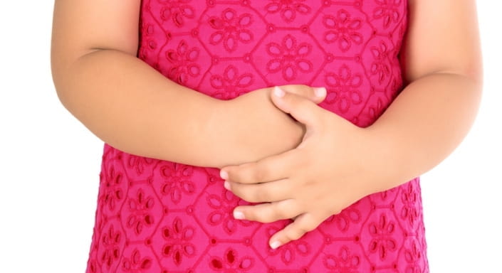
Figure 1. Abdominal Pain. (Johns, C., 2018)
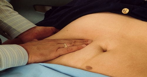
Figure 2 . Examination of Abdomen. (Kappan, S., 2018)
IMAGES
VIDEO
COMMENTS
A CASE STUDY ON CHOLELITHIASIS - Free download as Word Doc (.doc / .docx), PDF File (.pdf), Text File (.txt) or read online for free.
Case Study on Cholelithiasis - Free download as PDF File (.pdf) or read online for free. Surgical case presentation
Cholelithiasis Case Study - Free download as Word Doc (.doc), PDF File (.pdf), Text File (.txt) or read online for free. must be bsn students
Abstract-- Objective-- To describe the case about cholelithiasis. Clinical presentation and interventions-- A 45 year old female was visited tertiary care hospital with complain of right upper abdominal pain, nausea and vomiting .Her total bilirubin level was 3.5 mg/dl. The doctor advised hercholecystectomy.
However, gallstones can linger in the gallbladder for years without causing acute cholecystitis. Cholelithiasis often results in chronic inflammation of the gallbladder (chronic cholecystitis). Of note, elevated inflammatory markers and as shown in Figure 2, signs of gallbladder thickening suggest acute, as opposed to chronic cholecystitis.
Brown stones are a subtype of black gallstones, but contain more cholesterol and other fatty acids. Black stones are often innumerable, friable, and less than 1.0 cm. Brown gallstones tend to be soap-like and smooth. Mucin is a prominent matrix component involved with both stones' composition.
Gallstones (cholelithiasis) often obstruct the cystic duct and cause disease, and this patient does have several known risk factors for gallstones: age (>40 years old), female sex, obesity, diabetes, and oral contraceptive usage. Additional cholelithia-sis risk factors include rapid weight loss, pregnancy, and use of
1Guidelines Committee for Creating and Evaluating the "Evidence-Based Clinical Practice Guidelines for Cholelithiasis'', The Japanese Society of Gastroenterology, 6F Shimbashi i-MARK Building, 2-6-2 Shimbashi, Minato-ku, Tokyo 105-0004, Japan. 2Miyagi Medical Check-up Plaza, 1-6-9 Oroshi-machi, Wakabayashi-ku, Sendai, Miyagi 984-0015, Japan.
2. Pablo Becerra, a Valentina Becerra,a 2011 A case report on gallstones. Case Rep, 2011; 2(7): 228- 229. Published online, 2011 Jul 23. PMCID: PMC3199620 3. The study on clinical profile of patients with gallstones *1 Dr. Arvind Kumar Singh, 2 Dr. Sahjanand Prasad Singh International Journal of
The prevalence of cholelithiasis is approximately 20.5 million (6.3 million men and 14.2 million women) in the United States . Between 5% and 30% of patients with cholelithiasis develop concomitant choledocholithiasis . The diagnosis of choledocholithiasis is made based on the clinical signs and symptoms, results of liver function tests, and ...
cholelithiasis INTRODUTION: Gallstones (cholelithiasis) consists of deposits of digestive fluid that can form into a hardened stones in the gall bladder. The gall bladder is the small organ located just beneath the liver. The gall bladder holds the digestive fluids known as bile that is released into the small intestine.
325813139 a Case Study on Cholelithiasis - Free download as PDF File (.pdf), Text File (.txt) or read online for free. No significant family history of hereditary diseases.
Cholelithiasis: A Brief Review on Diagnostic Approach and Management in Clinical Practice. July 2020. DOI: 10.19080/ARGH.2020.15.555913. Authors: Febyan Febyan. Orthopaedic & Traumatology - Prof ...
2.1. Genes. A Swedish twin study showed that an inherited predisposition is responsible for 25% of the overall risk of developing gallstones [].Lithogenic genes 1 and 2 (Lith1 and Lith2), playing a role in liver cholesterol secretion and regulating bile flow, have been described in murine models.Their human counterparts are ABCG5 and ABCG8 [].A recent study of 214 children with cholelithiasis ...
Here we will discuss cases of classic symptomatology of cholecystitis and normal imaging studies, which were managed with cholecystectomy with complete resolution of symptoms. All final pathology reports confirmed the diagnosis of cholecystitis. CASES. Case 1: A 28-year-old-female presented with abdominal pain, nausea and vomiting. Physical ...
A-Case-Study-on-Cholelithiasis (1) - Read online for free. Hope maka help
a case presentation / study on cholelithiasis. Mar 7, 2020 • Download as PPTX, PDF •. 8 likes • 10,619 views. martinshaji. This is a case study prepared on cholecystitis (gall stones).For academic purpose of pharma D , and also for study aspects. Health & Medicine.
CASE STUDY ON Cholelithiasis - Free download as Word Doc (.doc / .docx), PDF File (.pdf), Text File (.txt) or view presentation slides online.
Case study on cholelithiasis. 1. Puspa Gauro MN 1st year 2068/10/. 2. To gain in-depth knowledge about the study subject/disease condition. To gain the confidence in handling such cases in future. To fulfill the partial course objective of M.N. curriculum. To share experience and knowledge to friends, juniors and seniors.
CASE STUDY OF CHOLELITHIASIS. I. Introduction. Cholelithiasis or gallstones are hardened deposits of digestive fluid that can form in your gallbladder. This can cause fever, chills, and yellowing of your skin and eyes if a stone blocks the duct to your pancreas, the organ may become inflamed wherein your gallbladder might burst, or rupture, if ...
Cholelithiasis Case Study - Free download as Word Doc (.doc / .docx), PDF File (.pdf), Text File (.txt) or read online for free. A CASE STUDY on cholelithiasis. Not mine and i don't own right but i thought it would be useful to nursing students ! A CASE STUDY on cholelithiasis. Not mine and i don't own right but i thought it would be useful to ...
Chronic cholecystitis or symptomatic gallbladder is a prolonged mechanical or functional disorder of abnormal gallbladder emptying. Most of the patients have recurrent pain attacks (acute biliary colic), but when pain lasts more than 24 hours, it requires urgent surgical intervention (acute cholecystitis). The length of a fully distended gallbladder is about 7 to 10 cm. We report a case ...
Patient Case Presentation. Mrs. G.B. is a 38 year old female who presents to the emergency department with complaints of severe abdominal pain. G.B reports that she has had similar pain intermittently over the past week, however, tonight her pain has become constant and unbearable. She reports that the pain usually starts on the right side of ...