Henry Gustav Molaison: The Curious Case of Patient H.M.
Erin Heaning
Clinical Safety Strategist at Bristol Myers Squibb
Psychology Graduate, Princeton University
Erin Heaning, a holder of a BA (Hons) in Psychology from Princeton University, has experienced as a research assistant at the Princeton Baby Lab.
Learn about our Editorial Process
Saul Mcleod, PhD
Editor-in-Chief for Simply Psychology
BSc (Hons) Psychology, MRes, PhD, University of Manchester
Saul Mcleod, PhD., is a qualified psychology teacher with over 18 years of experience in further and higher education. He has been published in peer-reviewed journals, including the Journal of Clinical Psychology.
On This Page:
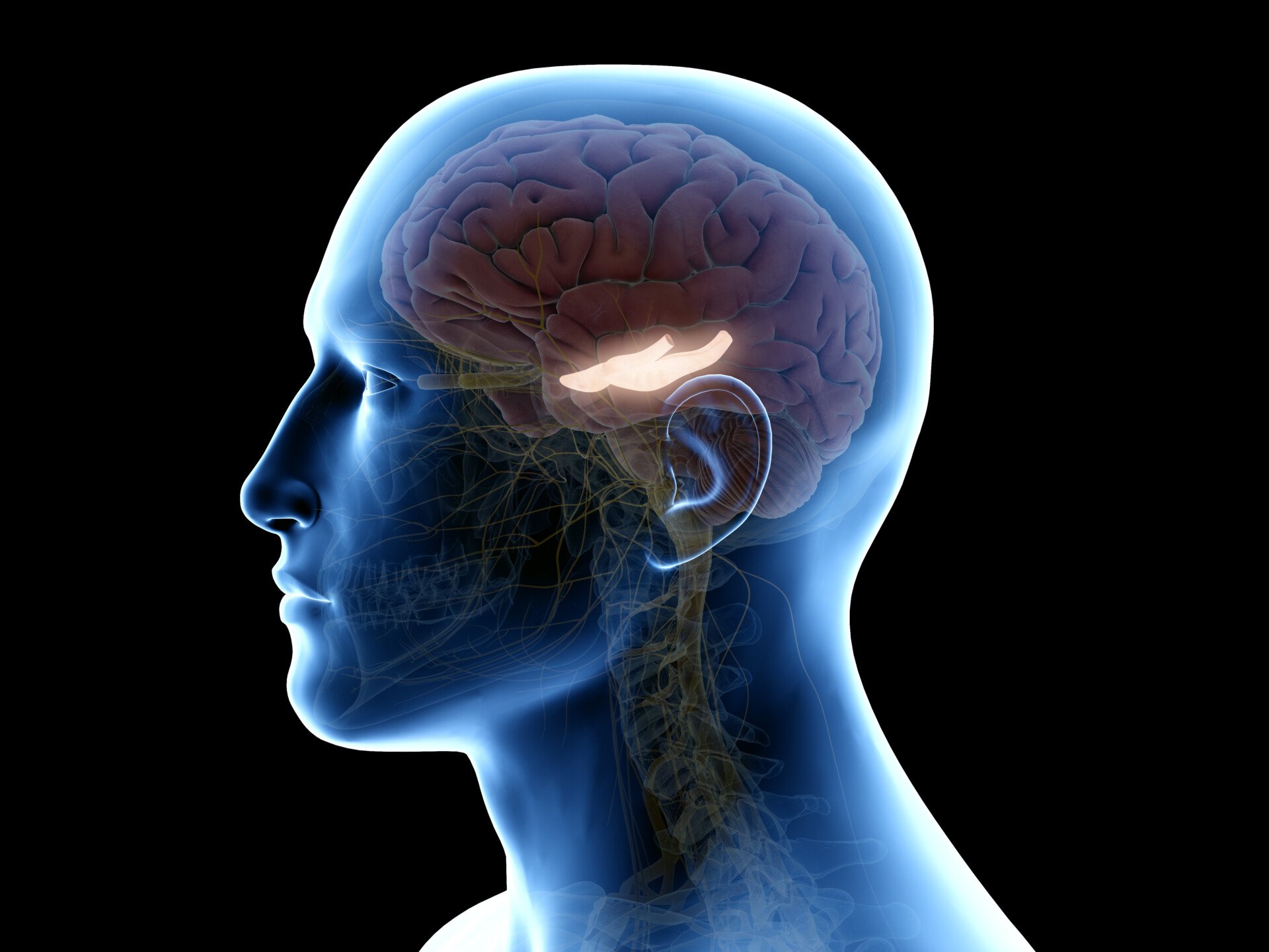
Henry Gustav Molaison, known as Patient H.M., is a landmark case study in psychology. After a surgery to alleviate severe epilepsy, which removed large portions of his hippocampus , he was left with anterograde amnesia , unable to form new explicit memories , thus offering crucial insights into the role of the hippocampus in memory formation.
- Henry Gustav Molaison (often referred to as H.M.) is a famous case of anterograde and retrograde amnesia in psychology.
- H. M. underwent brain surgery to remove his hippocampus and amygdala to control his seizures. As a result of his surgery, H.M.’s seizures decreased, but he could no longer form new memories or remember the prior 11 years of his life.
- He lost his ability to form many types of new memories (anterograde amnesia), such as new facts or faces, and the surgery also caused retrograde amnesia as he was able to recall childhood events but lost the ability to recall experiences a few years before his surgery.
- The case of H.M. and his life-long participation in studies gave researchers valuable insight into how memory functions and is organized in the brain. He is considered one of the most studied medical and psychological history cases.

Who is H.M.?
Henry Gustav Molaison, or “H.M” as he is commonly referred to by psychology and neuroscience textbooks, lost his memory on an operating table in 1953.
For years before his neurosurgery, H.M. suffered from epileptic seizures believed to be caused by a bicycle accident that occurred in his childhood. The seizures started out as minor at age ten, but they developed in severity when H.M. was a teenager.
Continuing to worsen in severity throughout his young adulthood, H.M. was eventually too disabled to work. Throughout this period, treatments continued to turn out unsuccessful, and epilepsy proved a major handicap and strain on H.M.’s quality of life.
And so, at age 27, H.M. agreed to undergo a radical surgery that would involve removing a part of his brain called the hippocampus — the region believed to be the source of his epileptic seizures (Squire, 2009).
For epilepsy patients, brain resection surgery refers to removing small portions of brain tissue responsible for causing seizures. Although resection is still a surgical procedure used today to treat epilepsy, the use of lasers and detailed brain scans help ensure valuable brain regions are not impacted.
In 1953, H.M.’s neurosurgeon did not have these tools, nor was he or the rest of the scientific or medical community fully aware of the true function of the hippocampus and its specific role in memory. In one regard, the surgery was successful, as H.M. did, in fact, experience fewer seizures.
However, family and doctors soon noticed he also suffered from severe amnesia, which persisted well past when he should have recovered. In addition to struggling to remember the years leading up to his surgery, H.M. also had gaps in his memory of the 11 years prior.
Furthermore, he lacked the ability to form new memories — causing him to perpetually live an existence of moment-to-moment forgetfulness for decades to come.
In one famous quote, he famously and somberly described his state as “like waking from a dream…. every day is alone in itself” (Squire et al., 2009).
H.M. soon became a major case study of interest for psychologists and neuroscientists who studied his memory deficits and cognitive abilities to better understand the hippocampus and its function.
When H.M. died on December 2, 2008, at the age of 82, he left behind a lifelong legacy of scientific contribution.
Surgical Procedure
Neurosurgeon William Beecher Scoville performed H.M.’s surgery in Hartford, Connecticut, in August 1953 when H.M. was 27 years old.
During the procedure, Scoville removed parts of H.M.’s temporal lobe which refers to the portion of the brain that sits behind both ears and is associated with auditory and memory processing.
More specifically, the surgery involved what was called a “partial medial temporal lobe resection” (Scoville & Milner, 1957). In this resection, Scoville removed 8 cm of brain tissue from the hippocampus — a seahorse-shaped structure located deep in the temporal lobe .
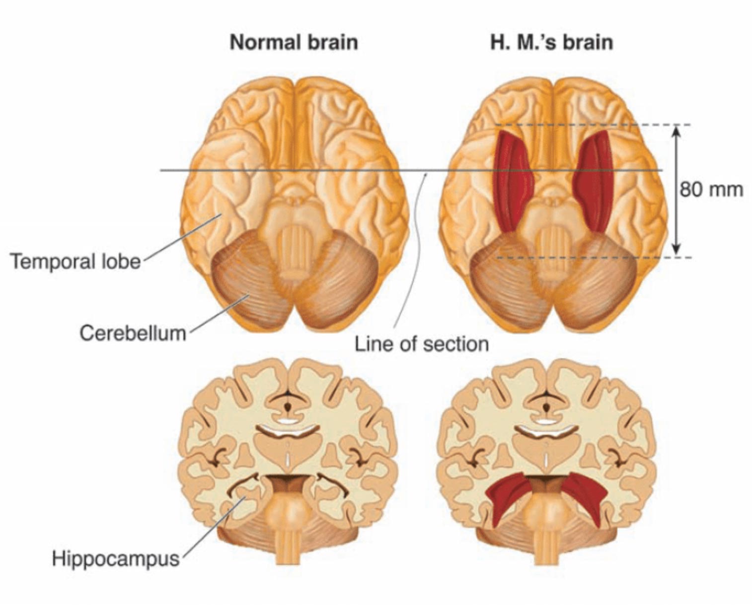
Bilateral resection of the anterior temporal lobe in patient HM.
Further research conducted after this removal showed Scoville also probably destroyed the brain structures known as the “uncus” (theorized to play a role in the sense of smell and forming new memories) and the “amygdala” (theorized to play a crucial role in controlling our emotional responses such as fear and sadness).
As previously mentioned, the removal surgery partially reduced H.M.’s seizures; however, he also lost the ability to form new memories.
At the time, Scoville’s experimental procedure had previously only been performed on patients with psychosis, so H.M. was the first epileptic patient and showed no sign of mental illness. In the original case study of H.M., which is discussed in further detail below, nine of Scoville’s patients from this experimental surgery were described.
However, because these patients had disorders such as schizophrenia, their symptoms were not removed after surgery. In this regard, H.M. was the only patient with “clean” amnesia along with no other apparent mental problems.
H.M’s Amnesia
H.M.’s apparent amnesia after waking from surgery presented in multiple forms. For starters, H.M. suffered from retrograde amnesia for the 11-year period prior to his surgery.
Retrograde describes amnesia, where you can’t recall memories that were formed before the event that caused the amnesia. Important to note, current research theorizes that H.M.’s retrograde amnesia was not actually caused by the loss of his hippocampus, but rather from a combination of antiepileptic drugs and frequent seizures prior to his surgery (Shrader 2012).
In contrast, H.M.’s inability to form new memories after his operation, known as anterograde amnesia, was the result of the loss of the hippocampus.
This meant that H.M. could not learn new words, facts, or faces after his surgery, and he would even forget who he was talking to the moment he walked away.
However, H.M. could perform tasks, and he could even perform those tasks easier after practice. This important finding represented a major scientific discovery when it comes to memory and the hippocampus. The memory that H.M. was missing in his life included the recall of facts, life events, and other experiences.
This type of long-term memory is referred to as “explicit” or “ declarative ” memories and they require conscious thinking.
In contrast, H.M.’s ability to improve in tasks after practice (even if he didn’t recall that practice) showed his “implicit” or “ procedural ” memory remained intact (Scoville & Milner, 1957). This type of long-term memory is unconscious, and examples include riding a bike, brushing your teeth, or typing on a keyboard.
Most importantly, after removing his hippocampus, H.M. lost his explicit memory but not his implicit memory — establishing that implicit memory must be controlled by some other area of the brain and not the hippocampus.
After the severity of the side effects of H.M.’s operation became clear, H.M. was referred to neurosurgeon Dr. Wilder Penfield and neuropsychologist Dr. Brenda Milner of Montreal Neurological Institute (MNI) for further testing.
As discussed, H.M. was not the only patient who underwent this experimental surgery, but he was the only non-psychotic patient with such a degree of memory impairment. As a result, he became a major study and interest for Milner and the rest of the scientific community.
Since Penfield and Milner had already been conducting memory experiments on other patients at the time, they quickly realized H.M.’s “dense amnesia, intact intelligence, and precise neurosurgical lesions made him a perfect experimental subject” (Shrader 2012).
Milner continued to conduct cognitive testing on H.M. for the next fifty years, primarily at the Massachusetts Institute of Technology (MIT). Her longitudinal case study of H.M.’s amnesia quickly became a sensation and is still one of the most widely-cited psychology studies.
In publishing her work, she protected Henry’s identity by first referring to him as the patient H.M. (Shrader 2012).
In the famous “star tracing task,” Milner tested if H.M.’s procedural memory was affected by the removal of the hippocampus during surgery.
In this task, H.M. had to trace an outline of a star, but he could only trace the star based on the mirrored reflection. H.M. then repeated this task once a day over a period of multiple days.
Over the course of these multiple days, Milner observed that H.M. performed the test faster and with fewer errors after continued practice. Although each time he performed the task, he had no memory of having participated in the task before, his performance improved immensely (Shrader 2012).
As this task showed, H.M. had lost his declarative/explicit memory, but his unconscious procedural/implicit memory remained intact. Given the damage to his hippocampus in surgery, researchers concluded from tasks such as these that the hippocampus must play a role in declarative but not procedural memory.
Therefore, procedural memory must be localized somewhere else in the brain and not in the hippocampus.
H.M’s Legacy
Milner’s and hundreds of other researchers’ work with H.M. established fundamental principles about how memory functions and is organized in the brain.
Without the contribution of H.M. in volunteering the study of his mind to science, our knowledge today regarding the separation of memory function in the brain would certainly not be as strong.
Until H.M.’s watershed surgery, it was not known that the hippocampus was essential for making memories and that if we lost this valuable part of our brain, we would be forced to live only in the moment-to-moment constraints of our short-term memory .
Once this was realized, the findings regarding H.M. were widely publicized so that this operation to remove the hippocampus would never be done again (Shrader 2012).
H.M.’s case study represents a historical time period for neuroscience in which most brain research and findings were the result of brain dissections, lesioning certain sections, and seeing how different experimental procedures impacted different patients.
Therefore, it is paramount we recognize the contribution of patients like H.M., who underwent these dangerous operations in the mid-twentieth century and then went on to allow researchers to study them for the rest of their lives.
Even after his death, H.M. donated his brain to science. Researchers then took his unique brain, froze it, and then in a 53-hour procedure, sliced it into 2,401 slices which were then individually photographed and digitized as a three-dimensional map.
Through this map, H.M.’s brain could be preserved for posterity (Wb et al., 2014). As neuroscience researcher Suzanne Corkin once said it best, “H.M. was a pleasant, engaging, docile man with a keen sense of humor, who knew he had a poor memory but accepted his fate.
There was a man behind the data. Henry often told me that he hoped that research into his condition would help others live better lives. He would have been proud to know how much his tragedy has benefitted science and medicine” (Corkin, 2014).
Corkin, S. (2014). Permanent present tense: The man with no memory and what he taught the world. Penguin Books.
Hardt, O., Einarsson, E. Ö., & Nader, K. (2010). A bridge over troubled water: Reconsolidation as a link between cognitive and neuroscientific memory research traditions. Annual Review of Psychology, 61, 141–167.
Scoville, W. B., & Milner, B. (1957). Loss of recent memory after bilateral hippocampal lesions . Journal of neurology, neurosurgery, and psychiatry, 20 (1), 11.
Shrader, J. (2012, January). HM, the man with no memory | Psychology Today. Retrieved from, https://www.psychologytoday.com/us/blog/trouble-in-mind/201201/hm-the-man-no-memory
Squire, L. R. (2009). The legacy of patient H. M. for neuroscience . Neuron, 61 , 6–9.

HM, the Man with No Memory
Henry molaison (hm) taught us about memory by losing his..
Posted January 16, 2012 | Reviewed by Jessica Schrader
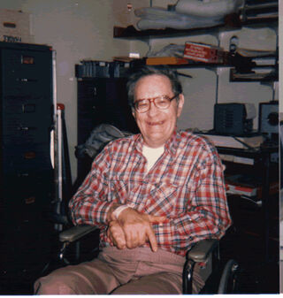
Henry Molaison, known by thousands of psychology students as "HM," lost his memory on an operating table in a hospital in Hartford in August 1953. He was 27 years old and had suffered from epileptic seizures for many years.
William Beecher Scoville, a Hartford neurosurgeon , stood above an awake Henry and skilfully suctioned out the seahorse-shaped brain structure called the hippocampus that lay within each temporal lobe. Henry would have been drowsy and probably didn't notice his memory vanishing as the operation proceeded.
The operation was successful in that it significantly reduced Henry's seizures, but it left him with a dense memory loss. When Scoville realized his patient had become amnesic, he referred him to the eminent neurosurgeon Dr. Wilder Penfield and neuropsychologist Dr. Brenda Milner of Montreal Neurological Institute (MNI), who assessed him in detail. Up until then, it had not been known that the hippocampus was essential for making memories, and that if we lose both of them we will suffer a global amnesia. Once this was realized, the findings were widely publicized so that this operation to remove both hippocampi would never be done again.
Penfield and Milner had already been conducting memory experiments on other patients and they quickly realized that Henry's dense amnesia, his intact intelligence , and the precise neurosurgical lesions made him the perfect experimental subject. For 55 years, Henry participated in numerous experiments, primarily at Massachusetts Institute of Technology (MIT), where Professor Suzanne Corkin and her team of neuropsychologists assessed him.
Access to Henry was carefully restricted to less than 100 researchers (I was honored to be one of them), but the MNI and MIT studies on HM taught us much of what we know about memory. Of course, many other patients with memory impairments have since been studied, including a small number with amnesias almost as dense as Henry's, but it is to him we owe the greatest debt. His name (or initials!) has been mentioned in almost 12,000 journal articles, making him the most studied case in medical or psychological history. Henry died on December 2, 2008, at the age of 82. Until then, he was known to the world only as "HM," but on his death his name was revealed. A man with no memory is vulnerable, and his initials had been used while he lived in order to protect his identity .
Henry's memory loss was far from simple. Not only could he make no new conscious memories after his operation, he also suffered a retrograde memory loss (a loss of memories prior to brain damage) for an 11-year period before his surgery. It is not clear why this is so, although it is thought this is not because of his loss of the hippocampi on both sides of his brain. More likely it is a combination of his being on large doses of antiepileptic drugs and his frequent seizures prior to his surgery. His global amnesia for new material was the result of the loss of both hippocampi, and meant that he could not learn new words, songs or faces after his surgery, forgot who he was talking to as soon as he turned away, didn't know how old he was or if his parents were alive or dead, and never again clearly remembered an event, such as his birthday party, or who the current president of the United States was.
In contrast, he did retain the ability to learn some new motor skills, such as becoming faster at drawing a path through a picture of a maze, or learning to use a walking frame when he sprained his ankle, but this learning was at a subconscious level. He had no conscious memory that he had ever seen or done the maze test before, or used the walking frame previously.
We measure time by our memories, and thus for Henry, it was as if time stopped when he was 16 years old, 11 years before his surgery. Because his intelligence in other non-memory areas remained normal, he was an excellent experimental participant. He was also a very happy and friendly person and always a delight to be with and to assess. He never seemed to get tired of doing what most people would think of as tedious memory tests, because they were always new to him! When he was at MIT, between test sessions he would often sit doing crossword puzzles, and he could do the same ones again and again if the words were erased, as to him it was new each time.
Henry gave science the ultimate gift: his memory. Thousands of people who have suffered brain damage, whether through accident, disease or a genetic quirk, have given similar gifts to science by agreeing to participate in psychological, neuropsychological, psychiatric and medical studies and experiments, and in some cases by gifting their brains to science after their deaths. Our knowledge of brain disease and how the normal mind works would be greatly diminished if it were not for the generosity of these people and their families (who are frequently also involved in interviews, as well as transporting the "patient" back and forth to the psychology laboratory). After Henry's death, his brain was dissected into 2,000 slices and digitized as a three-dimensional brain map that could be searched by zooming in from the whole brain to individual neurons. Thus, his tragically unique brain has been preserved for posterity.

Jenni Ogden, Ph.D. , clinical neuropsychologist and author of Trouble in Mind, taught at the University of Auckland.
- Find a Therapist
- Find a Treatment Center
- Find a Psychiatrist
- Find a Support Group
- Find Teletherapy
- United States
- Brooklyn, NY
- Chicago, IL
- Houston, TX
- Los Angeles, CA
- New York, NY
- Portland, OR
- San Diego, CA
- San Francisco, CA
- Seattle, WA
- Washington, DC
- Asperger's
- Bipolar Disorder
- Chronic Pain
- Eating Disorders
- Passive Aggression
- Personality
- Goal Setting
- Positive Psychology
- Stopping Smoking
- Low Sexual Desire
- Relationships
- Child Development
- Therapy Center NEW
- Diagnosis Dictionary
- Types of Therapy

Understanding what emotional intelligence looks like and the steps needed to improve it could light a path to a more emotionally adept world.
- Coronavirus Disease 2019
- Affective Forecasting
- Neuroscience
An official website of the United States government
The .gov means it’s official. Federal government websites often end in .gov or .mil. Before sharing sensitive information, make sure you’re on a federal government site.
The site is secure. The https:// ensures that you are connecting to the official website and that any information you provide is encrypted and transmitted securely.
- Publications
- Account settings
Preview improvements coming to the PMC website in October 2024. Learn More or Try it out now .
- Advanced Search
- Journal List
- HHS Author Manuscripts

The Legacy of Patient H.M. for Neuroscience
Larry r. squire.
1 Veterans Affairs Healthcare System, San Diego, CA 92161, USA
2 Departments of Psychiatry, Neurosciences, and Psychology, University of California, San Diego, La Jolla, CA 92093, USA
H.M. is probably the best known single patient in the history of neuroscience. His severe memory impairment, which resulted from experimental neurosurgery to control seizures, was the subject of study for five decades until his death in December 2008. Work with H.M. established fundamental principles about how memory functions are organized in the brain.
In 1952, Brenda Milner was completing her doctoral research at McGill University under the direction of Donald Hebb. At about this time, she encountered two patients (P.B. and F.C.) who had become severely amnesic following unilateral removal of the medial structures of the left temporal lobe for the treatment of epileptic seizures ( Penfield and Milner, 1958 ). This unfortunate outcome was entirely unexpected, and it was proposed that in each case there had been a preexistent, but unsuspected, atrophic lesion in the medial temporal lobe of the opposite hemisphere. In that way, the unilateral surgery would have resulted in a bilateral lesion, an idea that was confirmed at autopsy some years later for patient P.B. After the two cases were presented at the 1955 meeting of the American Neurological Association, Wilder Penfield (the neurosurgeon in both cases) received a call from William Scoville, a neurosurgeon in Hartford, Connecticut. Scoville told Penfield that he had seen a similar memory impairment in one of his own patients (H.M.) in whom he had carried out a bilateral medial temporal lobe resection in an attempt to control epileptic seizures. As a result of this conversation, Brenda Milner was invited to travel to Hartford to study H.M.
H.M. had been knocked down by a bicycle at the age of 7, began to have minor seizures at age 10, and had major seizures after age 16. (The age of the bicycle accident is given as 9 in some reports; for clarification see Corkin, 1984 .) He worked for a time on an assembly line but, finally, in 1953 at the age of 27 he had become so incapacitated by his seizures, despite high doses of anticonvulsant medication, that he could not work or lead a normal life. Scoville offered H.M. an experimental procedure that he had carried out previously in psychotic patients, and the surgery was then performed with the approval of the patient and his family.
When Milner first visited H.M., she saw that the epilepsy was now controlled but that his memory impairment was even more severe than in Penfield’s two patients, P.B. and F.C. What she observed was someone who forgot daily events nearly as fast as they occurred, apparently in the absence of any general intellectual loss or perceptual disorder. He underestimated his own age, apologized for forgetting the names of persons to whom he had just been introduced, and described his state as “like waking from a dream ... every day is alone in itself...” ( Milner et al., 1968 , p. 217).
The first observations of H.M., and the results of formal testing, were reported a few years later ( Scoville and Milner, 1957 ). This publication became one of the most cited papers in neuroscience (nearly 2500 citations) and is still cited with high frequency. H.M. continued to be studied for five decades, principally by Brenda Milner, her former student Suzanne Corkin, and their colleagues ( Corkin, 1984 , 2002 ; Milner et al., 1968 ). He died on December 2, 2008, at the age of 82. It can be said that the early descriptions of H.M. inaugurated the modern era of memory research. Before H.M., due particularly to the influence of Karl Lashley, memory functions were thought to be widely distributed in the cortex and to be integrated with intellectual and perceptual functions. The findings from H.M. established the fundamental principle that memory is a distinct cerebral function, separable from other perceptual and cognitive abilities, and identified the medial aspect of the temporal lobe as important for memory. The implication was that the brain has to some extent separated its perceptual and intellectual functions from its capacity to lay down in memory the records that ordinarily result from engaging in perceptual and intellectual work.
The Medial Temporal Lobe Memory System
The early paper is sometimes cited incorrectly as evidence that the hippocampus is important for memory, but this particular point could not of course be established from a lesion that, by the surgeon’s description, included the hippocampus, amygdala, and the adjacent parahippocampal gyrus. As Milner subsequently wrote, “Despite the use of the word ‘hippocampal’ in the titles of my papers with Scoville and Penfield, I have never claimed that the memory loss was solely attributable to the hippocampal lesions” ( Milner, 1998 ). Indeed, the original paper ends, quite appropriately, with the statement:
It is concluded that the anterior hippocampus and hippocampal gyrus, either separately or together, are critically concerned in the retention of current experience. It is not known whether the amygdala plays any part in this mechanism, since the hippocampal complex has not been removed alone, but always together with uncus and amygdala. ( Scoville and Milner, 1957 , p. 21).
The findings from H.M. were initially met with some resistance, especially because of the difficulty for many years of demonstrating anything resembling his impairment in the experimental animal. Efforts to establish an animal model in fact began almost immediately when Scoville himself came to Montreal and did the same surgery in monkeys that he had done with H.M. But these monkeys and others with medial temporal lesions seemed able to learn tasks that H.M. could not learn. Only much later did it become understood that apparently similar tasks can be learned in different ways by humans and monkeys. For example, the visual discrimination task, which is learned gradually by the monkey over hundreds of trials, proved to involve what one would now call habit learning. In the monkey, this kind of learning depends on the basal ganglia, not the medial temporal lobe. Eventually, tasks were developed for the monkey that were exquisitely sensitive to medial temporal lobe lesions (for example, the one-trial, delayed nonmatching to sample task), and an animal model of human memory impairment thereby became available ( Mishkin, 1978 ).
Cumulative work with the animal model over the next decade, together with neuroanatomical studies, succeeded in identifying the anatomical components of what is now termed the medial temporal lobe memory system ( Squire and Zola-Morgan, 1991 ): the hippocampus and the adjacent perirhinal, entorhinal, and parahippocampal cortices that make up much of the parahippocampal gyrus. This information showed which structures within H.M.’s large lesion were important for understanding his impairment and, more broadly, what structures are important for memory. A few years later, an improved description of H.M.’s lesion was obtained with magnetic resonance imaging (MRI) ( Corkin et al., 1997 ). MRI had been delayed because of concerns that clips placed on the dura during surgery made H.M. ineligible for imaging. However, thorough inquiry revealed that the dural clips constituted no risk.
At this juncture, several points became clear. First, H.M.’s lesion was less extensive than described originally by the surgeon in that it extended a little more than 5 cm caudally from the temporal pole (not 8 cm). As a result the posterior parahippocampal gyrus was largely spared (specifically, the parahippocampal cortex or what in the monkey is termed area TH TF). Second, the reason that H.M.’s memory impairment was so severe was that the bilateral damage included the parahippocampal gyrus (anteriorly) and was not restricted to the hippocampus. Damage limited to the hippocampus causes significant memory impairment but considerably less impairment than in H.M. Third, memory impairment more severe than H.M.’s could now be understood, as when the damage includes the structures damaged in H.M. but also extends far enough posteriorly to involve the parahippocampal cortex (patients E.P. and G.P.; Kirwan et al., 2008 ).
In the early years, the anatomy of the medial temporal lobe was poorly understood, and terms like hippocampal zone and hippocampal complex were often used to identify the area of damage. With the elucidation of the boundaries and connectivity of the structures adjacent to the hippocampus and the discovery that these structures are important for memory, vague terms like hippocampal complex became unnecessary (though one can still find them in contemporary writing). It is now possible to achieve careful descriptions based on anatomical measurement and modern terminology.
H.M. not only motivated the development of an animal model of human memory impairment and the subsequent delineation of the medial temporal lobe memory system. As described next, the study of H.M. also led to fundamental insights into the function of the medial temporal lobe and the larger matter of how memory is organized in the brain.
Immediate Memory and Long-Term Memory
H.M.’s intact intellectual and perceptual functions, and similar findings in other patients with large medial temporal lesions, have been well documented. A key additional finding was that H.M. had a remarkable capacity for sustained attention, including the ability to retain information for a period of time after it was presented. Thus, he could carry on a conversation, and he exhibited an intact digit span (i.e., the ability to repeat back a string of six or seven digits). Indeed, information remained available so long as it could be actively maintained by rehearsal. For example, H.M. could retain a three-digit number for as long as 15 min by continuous rehearsal, organizing the digits according to an elaborate mnemonic scheme. Yet when his attention was diverted to a new topic, he forgot the whole event. In contrast, when the material was not easy to rehearse (in the case of nonverbal stimuli like faces or designs), information slipped away in less than a minute. These findings supported a fundamental distinction between immediate memory and long-term memory (what William James termed primary memory and secondary memory). Primary memory [immediate memory]
...comes to us as belonging to the rearward portion of the present space of time, and not to the genuine past ( James, 1890 , p. 647).
Secondary memory [long-term memory] is quite different.
An object which has been recollected. is one which has been absent from consciousness altogether, and now revives anew. It is brought back, recalled, fished up, so to speak, from a reservoir in which, with countless other objects, it lay buried and lost from view. ( James, 1890 , p. 648).
Notably, time is not the key factor that determines how long patients like H.M. can retain information in memory. The relevant factors are the capacity of immediate memory and attention, i.e., the amount of material that can be held in mind and how successfully it can be rehearsed. The work with H.M. demonstrated that the psychological distinction between immediate memory and long-term memory is a prominent feature of how the brain has organized its memory functions.
Multiple Memory Systems
Perhaps the most unexpected discovery about H.M., given his profound and global memory impairment, came when Brenda Milner tested his ability to acquire a visuomotor skill ( Milner, 1962 ). H.M. was shown a five-pointed star, with a double contour, and asked to trace its outline with a pencil, but in a condition when he could only see his hand and the star as reflected in a mirror. H.M. acquired this mirror-drawing skill during ten trials and exhibited excellent retention across 3 days. Yet at the end of testing, he had no recollection of having done the task before. This demonstration provided the first hint that there was more than one kind of memory in the brain and suggested that some kinds of memory (motor skills) must lie outside the province of the medial temporal lobe.
For a time, it was rather thought that motor skills were a special case and that all the rest of memory is impaired in patients like H.M. Later it became appreciated that motor skills are but a subset of a larger domain of skill-like abilities, all of which are preserved in amnesia. The demonstration of a fully preserved ability to learn the perceptual skill of mirror reading suggested a distinction between two broad classes of knowledge: declarative and procedural ( Cohen and Squire, 1980 ). Declarative memory is what is meant when the term “memory” is used in everyday language, i.e., conscious knowledge of facts and events. Procedural memory refers to skill-based knowledge that develops gradually but with little ability to report what is being learned.
In the years that followed, other preserved learning abilities began to be reported for amnesic patients, and the perspective shifted to a framework that accommodated multiple (i.e., more than two) memory systems. As Endel Tulving wrote:
But even if we accept the broad division of memory into procedural and propositional forms ... there are phenomena that do not seem to fit readily into such a taxonomy ( Tulving et al., 1982 , p.336).
Subsequently, the terms declarative and nondeclarative were introduced with the idea that declarative memory refers to the kind of memory that is impaired in H.M. and is dependent on the medial temporal lobe. Nondeclarative memory is an umbrella term referring to additional memory systems. These include systems that support skill learning, habit learning, simple conditioning, emotional learning, as well as priming and perceptual learning. The structures with special importance for these kinds of memory include the basal ganglia, the cerebellum, the amygdala, and the neocortex. The starting point for these developments was the early discovery that motor skill learning was preserved in H.M. This finding revealed that memory is not a single faculty of the mind and led ultimately to the identification of the multiple memory systems of the mammalian brain.
Remote Memory
H.M.’s memory impairment has generally been taken as reflecting a failure to convert transient, immediate memory into stable long-term memory. A key insight about the organization of memory, and medial temporal lobe function, came with a consideration of his capacity to remember information that he had acquired before his surgery. The first exploration of this issue with formal tests asked H.M. to recognize faces of persons who had become famous in different decades, 1920-1970 ( Marslen-Wilson and Teuber, 1975 ). As expected, H.M. was severely impaired at recognizing faces from his postmorbid period (the 1950s and 1960s), but he performed as well as or better than age-matched controls at recognizing faces of persons who were in the news before his surgery. This important finding implied that the medial temporal lobe is not the ultimate storage site for previously acquired knowledge. The early descriptions of H.M. conform to this view. Thus, H.M. was described as having a partial loss of memory (retrograde amnesia) for the 3 years leading up to his surgery, with early memories “seemingly normal” ( Scoville and Milner, 1957 , p. 17). Similarly, about 10 years later it was remarked that there did not appear
to have been any change in H.M.’s capacity to recall remote events antedating his operation, such as incidents from his early school years, a high-school attachment, or jobs he had held in his late teens and early twenties ( Milner et al., 1968 , p. 216).
Subsequently, a particular interest developed in the status of autobiographical memories for unique events, which are specific to time and place, and methods were developed to assess the specificity and the detail with which such recollections could be reproduced. In the earliest efforts along these lines, as summarized by Suzanne Corkin ( Corkin, 1984 ), H.M. produced well-formed autobiographical memories, from age 16 years or younger. It was concluded that H.M’s remote memory impairment now extended back to 11 years before his surgery. The situation seemed to change further as H.M. aged. In an update prepared nearly 20 years later ( Corkin, 2002 ), H.M. (now 76 years old) was described as having memories of childhood, but his memories appeared more like remembered facts than like memories of specific episodes. It was also said that he could not narrate a single event that occurred at a specific time and place. Essentially the same conclusion was reached a few years later when new methods, intended to be particularly sensitive, were used to assess H.M.’s remote memory for autobiographical events ( Steinvorth et al., 2005 ). These later findings led to the proposal that, whatever might be the case for fact memory, autobiographical memories, i.e., memories that are specific to time and place, depend on the medial temporal lobe so long as the memories persist.
There are reasons to be cautious about this idea. In 2002-2003, new MRI scans of H.M. were obtained ( Salat et al., 2006 ). These scans documented a number of changes since his first MRI scans from 1992-1993 ( Corkin et al., 1997 ), including cortical thinning, subcortical atrophy, large amounts of abnormal white matter, and subcortical infarcts. These findings were thought to have appeared during the past decade, and they complicate the interpretation of neuropsychological data collected during the same time period. Another consideration is that remote memories could have been intact in the early years after surgery but then have faded with time because they could not be strengthened through rehearsal and relearning. In any case, the optimal time to assess the status of past memory is soon after the onset of memory impairment.
Other work has tended to support the earlier estimates that H.M.’s remote memories were intact. First, Penfield’s two patients described above, P.B. and F.C., were reported after their surgeries to have memory loss extending back a few months and 4 years, respectively, and intact memory from before that time ( Penfield and Milner, 1958 ). Second, methods like those used recently to assess H.M. have also been used to evaluate autobiographical memory in other patients, including patients like E.P. and G.P. who have very severe memory impairment ( Kirwan et al., 2008 ). In these cases, autobiographical recollection was impaired when memories were drawn from the recent past but fully intact when memories were drawn from the remote past.
Memory loss can sometimes extend back for decades in the case of large medial temporal lobe lesions (though additional damage to anterolateral temporal cortex may be important in this circumstance). In any case, memories from early life appear to be intact unless the damage extends well into the lateral temporal lobe or the frontal lobe. These findings are typically interpreted to mean that the structures damaged in H.M. are important for the formation of long-term memory and its maintenance for a period of time after learning. During this period gradual changes are thought to occur in neocortex (memory consolidation) that increase the complexity, distribution, and connectivity among multiple cortical regions. Eventually, memory can be supported by the neocortex and becomes independent of the medial temporal lobe. The surprising observation that H.M. had access to old memories, in the face of an inability to establish new ones, motivated an enormous body of work, both in humans and experimental animals, on the topic of remote memory and continues to stimulate discussion about the nature and significance of retrograde amnesia.
Perspective
H.M. was likely the most studied individual in the history of neuroscience. Interest in the case can be attributed to a number of factors, including the unusual purity and severity of the memory impairment, its stability, its well-described anatomical basis, and H.M.’s willingness to be studied. He was a quiet and courteous man with a sense of humor and insight into his condition. Speaking of his neurosurgeon, he once said, “What he learned about me helped others, and I’m glad about that.” ( Corkin, 2002 , p. 159).
An additional aspect of H.M.’s circumstance, which assured his eventual place in the history of neuroscience, was the fact that Brenda Milner was the young scientist who first studied him. She is a superb experimentalist with a strong conceptual orientation that allowed her to draw from her data deep insights about the organization of memory. Because he was the first well-studied patient with amnesia, H.M. became the yardstick against which other patients with memory impairment would be compared. It is now clear that his memory impairment was not absolute and that he was able to acquire significant new knowledge ( Corkin, 2002 ). Thus, memory impairment can be either more severe or less severe than in H.M. But the study of H.M. established key principles about how memory is organized that continue to guide the discipline.
ACKNOWLEDGMENTS
Supported by the Medical Research Service of the Department of Veterans Affairs, The National Institute of Mental Health (MH24600), and the Metro-politan Life Foundation. I thank Nicola Broadbent, Robert Clark, Christine Smith, Ryan Squire, and Wendy Suzuki for their helpful comments.
- Cohen N, Squire LR. Science. 1980; 210 :207–209. [ PubMed ] [ Google Scholar ]
- Corkin S. Semin. Neurol. 1984; 4 :249–259. [ Google Scholar ]
- Corkin S. Nat. Rev. Neurosci. 2002; 3 :153–160. [ PubMed ] [ Google Scholar ]
- Corkin S, Amaral DG, Gonzalez RG, Johnson KA, Hyman BT. J. Neurosci. 1997; 17 :3964–3979. [ PMC free article ] [ PubMed ] [ Google Scholar ]
- James W. Principles of Psychology. Dover Edition One. Holt; New York: 1890. [ Google Scholar ]
- Kirwan CB, Bayley PJ, Galván VV, Squire LR. Proc. Natl. Acad. Sci. USA. 2008; 105 :2676–2680. [ PMC free article ] [ PubMed ] [ Google Scholar ]
- Marslen-Wilson WD, Teuber HL. Neuropsychologia. 1975; 13 :353–364. [ PubMed ] [ Google Scholar ]
- Milner B. In: Physiologie de l’hippocampe. Passouant P, editor. Centre National de la Recherche Scientifique; Paris: 1962. pp. 257–272. [ Google Scholar ]
- Milner B. In: The History of Neuroscience in Autobiography. Squire LR, editor. Vol. 2. Academic Press; San Diego: 1998. pp. 276–305. [ Google Scholar ]
- Milner B, Corkin S, Teuber HL. Neuropsychologia. 1968; 6 :215–234. [ Google Scholar ]
- Mishkin M. Nature. 1978; 273 :297–298. [ PubMed ] [ Google Scholar ]
- Penfield W, Milner B. AMA Arch. Neurol. Psychiatry. 1958; 79 :475–497. [ PubMed ] [ Google Scholar ]
- Salat DH, van der Kouwe AJW, Tuch DS, Quinn BT, Fischl B, Dale AM, Corkin S. Hippocampus. 2006; 16 :936–945. [ PubMed ] [ Google Scholar ]
- Scoville WB, Milner B. J. Neurol. Neurosurg. Psychiatry. 1957; 20 :11–21. [ PMC free article ] [ PubMed ] [ Google Scholar ]
- Squire LR, Zola-Morgan S. Science. 1991; 253 :1380–1386. [ PubMed ] [ Google Scholar ]
- Steinvorth S, Levine B, Corkin S. Neuropsychologia. 2005; 43 :479–496. [ PubMed ] [ Google Scholar ]
- Tulving E, Schacter DL, Stark HA. J. Exp. Psychol. Learn. Mem. Cogn. 1982; 8 :336–342. [ Google Scholar ]
- Brain Development
- Childhood & Adolescence
- Diet & Lifestyle
- Emotions, Stress & Anxiety
- Learning & Memory
- Thinking & Awareness
- Alzheimer's & Dementia
- Childhood Disorders
- Immune System Disorders
- Mental Health
- Neurodegenerative Disorders
- Infectious Disease
- Neurological Disorders A-Z
- Body Systems
- Cells & Circuits
- Genes & Molecules
- The Arts & the Brain
- Law, Economics & Ethics
Neuroscience in the News
- Supporting Research
- Tech & the Brain
- Animals in Research
- BRAIN Initiative
- Meet the Researcher
- Neuro-technologies
- Tools & Techniques
- Core Concepts
- For Educators
- Ask an Expert
- The Brain Facts Book

The Curious Case of Patient H.M.
- Reviewed 28 Aug 2018
- Author Deborah Halber
- Source BrainFacts/SfN
On September 1, 1953, time stopped for Henry Molaison. For roughly 10 years, the 27-year-old had suffered severe seizures. By 1953, they were so debilitating he could no longer hold down his job as a motor winder on an assembly line. On September 1, Molaison allowed surgeons to remove a thumb-sized section of tissue from each side of his brain. It was an experimental procedure that he and his surgeons hoped would quell the seizures wracking his brain.
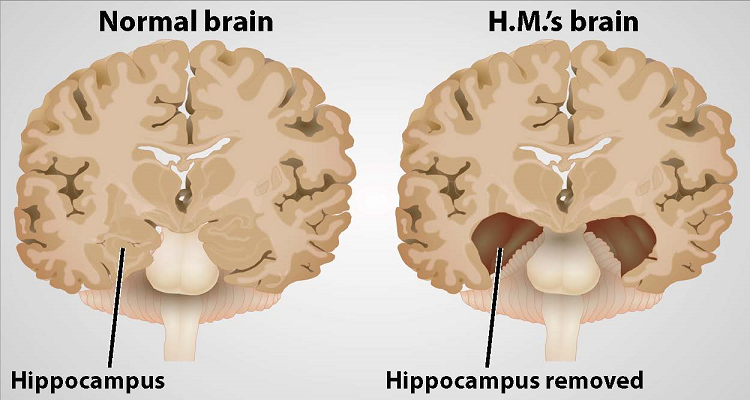
And, it worked. The seizures abated, but afterwards Molaison was left with permanent amnesia. He could remember some things — scenes from his childhood, some facts about his parents, and historical events that occurred before his surgery — but he was unable to form new memories. If he met someone who then left the room, within minutes he had no recollection of the person or their meeting.
What was a tragedy for Molaison led to one of the most significant turning points in 20th century brain science: the understanding that complex functions such as learning and memory are tied to discrete regions of the brain.
In 1955, scientists William Beecher Scoville and Brenda Milner began studying Molaison — referred to as H.M. to protect his privacy — and nine other patients who had undergone similar surgery. Only patients who had specific portions of their medial temporal lobes removed experienced memory problems. And, the more tissue removed, the more severe the memory impairment. The researchers noted patients’ amnesia was “curiously specific to the domain of recent memory.”
Scoville and Milner’s observations pointed to a particular structure within the medial temporal lobe that was necessary for normal memory — the hippocampus. Over the next five decades, neuroscientists studying Molaison learned that the hippocampus and adjacent regions transform our transient perceptions and awareness into memories that can last a lifetime.
For Molaison, this transformation could no longer take place. He experienced every aspect of his daily life — eating a meal, taking a walk — as a first. Yet his intellect, personality, and perception were intact, and he was able to acquire new motor skills. Over time, he became more proficient at tasks such as tracing patterns while watching his hand movements in a mirror, despite the fact that he could never recall performing the task before.
Studies of Molaison paved the way for further exploration of the brain networks encoding conscious and unconscious memories. Even after his death in 2008 at the age of 82, neuroscientists continue to learn from him.
This article was adapted from the 8th edition of Brain Facts by Deborah Halber.
About the Author

Deborah Halber
Deborah Halber is a Boston-based author, science writer and journalist. Her work has appeared in The Atlantic, Time.com, The Boston Globe, MIT Technology Review, Boston magazine, and university publications.
CONTENT PROVIDED BY
BrainFacts/SfN
What to Read Next

Also In Tools & Techniques
Popular articles on BrainFacts.org

Check out the latest news from the field.
Research & Discoveries
See how discoveries in the lab have improved human health.
BrainFacts Book
Download a copy of the newest edition of the book, Brain Facts: A Primer on the Brain and Nervous System.

SUPPORTING PARTNERS
- Privacy Policy
- Accessibility Policy
- Terms and Conditions
- Manage Cookies
Some pages on this website provide links that require Adobe Reader to view.

Encyclopedia of Clinical Neuropsychology pp 1629–1631 Cite as
H.M.; Also the Case of H.M., Molaison, Henry (1926–2008)
- Amy Alderson 4
- Reference work entry
- First Online: 01 January 2018
56 Accesses
Landmark Clinical, Scientific, and Professional Contributions
Investigations of Henry Molaison’s (HM) neurocognitive functioning following his surgical intervention have revolutionized our understanding of learning and memory processes. HM’s specific surgical intervention and associated cognitive impairments provided information not only about differential memory activities but also about the neural substrates involved in the mediation of these processes. Investigations of the alterations in HM’s memory led to a landmark paper by Brenda Milner, a psychologist working with HM, and William Scoville, his neurosurgeon. The paper, entitled Loss of recent memory after bilateral hippocampal lesions , was published in the Journal of Neurology, Neurosurgery, and Psychiatry in 1957; it brought about a sea change in the understanding of the neural substrates of memory. This paper has been cited more than 1800 times since its initial publication.
In this landmark paper, as well as in many others...
This is a preview of subscription content, log in via an institution .
Buying options
- Available as PDF
- Read on any device
- Instant download
- Own it forever
- Available as EPUB and PDF
- Durable hardcover edition
- Dispatched in 3 to 5 business days
- Free shipping worldwide - see info
Tax calculation will be finalised at checkout
Purchases are for personal use only
References and Readings
Corkin, S. (2002). Whats new with amnesic patient H.M.? Nature Reviews Neuroscience, 3 , 153–160.
Article PubMed Google Scholar
Ogden, J. A. (2005). Fractured minds . New York: Oxford University Press.
Google Scholar
Scoville, W. B. (1968). Amnesia after bilateral mesial temporal-lobe excision: Introduction to case H.M. Neuropsychologica, 6 , 211–213.
Article Google Scholar
Scoville, W. B., & Milner, B. (1957). Loss of recent memory after bilateral hippocampal lesions. Journal of Neurology, Neurosurgery, and Psychiatry, 20 , 11–21.
Article PubMed PubMed Central Google Scholar
Scoville, W. B., & Milner, B. (2000). Loss of recent memory after bilateral hippocampal lesions. Journal of Neuropsychiatry and Clinical Neuroscience, 12 (1), 103–113.
Download references
Author information
Authors and affiliations.
Department of Rehabilitation Medicine, Emory University, Atlanta, GA, USA
Amy Alderson
You can also search for this author in PubMed Google Scholar
Corresponding author
Correspondence to Amy Alderson .
Editor information
Editors and affiliations.
Department of Physical Medicine and Rehabilitation, Virginia Commonwealth University, Richmond, VA, USA
Jeffrey S. Kreutzer
Kessler Foundation, Pleasant Valley Way, West Orange, NJ, USA
John DeLuca
Independent Practice, Wynnewood, PA, USA
Bruce Caplan
Rights and permissions
Reprints and permissions
Copyright information
© 2018 Springer International Publishing AG, part of Springer Nature
About this entry
Cite this entry.
Alderson, A. (2018). H.M.; Also the Case of H.M., Molaison, Henry (1926–2008). In: Kreutzer, J.S., DeLuca, J., Caplan, B. (eds) Encyclopedia of Clinical Neuropsychology. Springer, Cham. https://doi.org/10.1007/978-3-319-57111-9_622
Download citation
DOI : https://doi.org/10.1007/978-3-319-57111-9_622
Published : 20 September 2018
Publisher Name : Springer, Cham
Print ISBN : 978-3-319-57110-2
Online ISBN : 978-3-319-57111-9
eBook Packages : Behavioral Science and Psychology Reference Module Humanities and Social Sciences Reference Module Business, Economics and Social Sciences
Share this entry
Anyone you share the following link with will be able to read this content:
Sorry, a shareable link is not currently available for this article.
Provided by the Springer Nature SharedIt content-sharing initiative
- Publish with us
Policies and ethics
- Find a journal
- Track your research
- Search Menu
- Advance articles
- Editor's Choice
- Continuing Education
- Author Guidelines
- Submission Site
- Open Access
- Why publish with this journal?
- About Archives of Clinical Neuropsychology
- About the National Academy of Neuropsychology
- Journals Career Network
- Editorial Board
- Advertising and Corporate Services
- Self-Archiving Policy
- Dispatch Dates
- Journals on Oxford Academic
- Books on Oxford Academic
Article Contents
Influential case studies, psychosurgery and asylums, temporal lobectomy, controversy, author notes.
- < Previous
Remembering H.M.: Review of “PATIENT H.M.: A Story of Memory, Madness, and Family Secrets”
- Article contents
- Figures & tables
- Supplementary Data
David W. Loring, Bruce Hermann, Remembering H.M.: Review of “PATIENT H.M.: A Story of Memory, Madness, and Family Secrets”, Archives of Clinical Neuropsychology , Volume 32, Issue 4, June 2017, Pages 501–505, https://doi.org/10.1093/arclin/acx041
- Permissions Icon Permissions
Although many influential case reports in neuropsychology exist ( Code, Wallesch, Joanette, & Lecours, 1996 ), there are certain patients who stand out because, based upon the historical zeitgeist in which their brain injuries occurred and the attention that those cases received, their neurobehavioral deficits and circumstances of their injury greatly altered our knowledge of brain-behavior relationships.
Among the most famous of these cases is Phineas Gage, the railroad foreman whose personality dramatically changed following frontal lobe injury in 1848 from an accidental explosion that thrust his tamping iron through his skull. Gage's survival after such a serious injury was a surprise, but Gage's contribution to clinical neuroscience was his significant personality change, aptly described by his physicians with the pithy observation, “Gage was no longer Gage” ( Macmillan, 2000 ). Although his personality changes were well documented soon after the accident, much of Gage's long-term outcome may have been exaggerated for entertainment value ( Macmillan & Lena, 2010 ). Thus, the lasting neurobehavioral effects of Gage's frontal lobe injury and how the deficits may have evolved over time remain clouded in the historical record due to the absence independent scientific characterization.
The second patient is Louis Victor Leborgne, whose expressive language disturbance from a left frontal lobe lesion was described in 1861 by the famous French neurologist Pierre Paul Broca. Monsieur Tan, as he was informally called because “tan tan” was his typical verbal output, retained his capacity to understand commands. The deficits of Monsieur Tan, supported by subsequent cases, demonstrated that language could be fractionated into different components associated with distinct brain regions, and that language was predominately a function of the left brain. Monsieur Tan's contribution, however, was in no small part due to Broca's distinguished reputation as a physician and scientist since localized language effects had been previously described by Jean-Baptiste Bouillard ( Sondhaus & Finger, 1988 ).
The third and most studied of these three cases is patient Henry Molaison (H.M.). H.M. suffered a dense and persistent anterograde amnesia following bilateral medial temporal lobectomy in 1953 to treat intractable epilepsy ( Scoville & Milner, 1957 ). His scientific fame derives from the dramatic demonstration of the critical role that the mesial temporal lobe structures play in learning and memory. Unlike Gage and Monsieur Tan, H.M.’s brain injury was iatrogenic, being an unanticipated adverse event associated with the surgical treatment of his epilepsy. Another important difference is that H.M.’s surgery injury occurred in what can broadly be considered to be the beginning of the modern era of neuroscience ( Shepherd, 2010 ). Thus, his cognitive abilities were subjected to formal characterization with extensive neuropsychological testing over five decades, providing a much richer characterization of his clinical semiology compared to Gage or Monsieur Tan.
H.M.’s amnesia framed how the neuroscience community would eventually conceptualize basic memory mechanisms, beginning with Brenda Milner's early demonstration that multiple memory systems exist such that declarative and procedural memory are readily dissociable ( Milner, 1965 ). Clinically, H.M.’s amnesia meaningfully influenced pre-operative epilepsy surgery protocols across the world. After several additional cases of post-surgical amnesia developed following unilateral temporal lobectomy, it was hypothesized that the functional reserve of the contralateral temporal lobe was insufficient to support the encoding of new memories following resection of the epileptogenic temporal lobe and mesial structures, and multiple methods for characterizing functional hippocampus status were developed ( Milner, 1975 ). What remains poorly reported in standard textbooks, however, is the historical context in which the decision to undergo epilepsy surgery was made, the blurring between experimental clinical techniques and informed consent, and the profound effects on H.M.’s quality of life.
To provide this broad historical context of H.M., Luke Dittrich has published PATIENT H.M.: A Story of Memory, Madness, and Family Secrets ( Dittrich, 2016a ). This is far from a narrative review of H.M.’s contributions to understanding memory, and it is also not a typical biography. However, as the grandson of William Beecher Scoville, MD, the neurosurgeon who performed H.M.’s operation and a prolific practitioner of psychosurgery, Dittrich provides a unique “insider” perspective and captivating description of that era's medical zeitgeist that could not be easily achieved without such a personal relationship. In fact, much of the book does not directly involve H.M.’s life story, but rather, the management of significant psychiatric disease prior to the development of neuroleptics.
Scoville's neurosurgical practice primarily involved surgery for psychiatric indications rather than epilepsy surgery. The early development of psychosurgery's goals is exemplified with a quote from the 19th century physician Dr. Gottleib Burckhardt, who resected undifferentiated brain areas, that illustrates the depersonalization of patients with psychiatric disease: “Mrs. B. has changed from a dangerous and excited demented person to a quiet demented one” (p. 79). It was in late 1935, after listening to the report of operations on two chimpanzees, that Egas Moniz oversaw the first in his series of approximately 20 frontal leucotomies/lobotomies. This series significantly influenced Walter Freeman (neurologist) and James Watts (neurosurgeon) who initially worked together performing prefrontal lobotomies. The distinct approaches to frontal lobotomy developed by Scoville and Freeman also provide a striking contrast in how to best decrease the institutional burden of psychiatric disease. Although Scoville is described as an adventurer who liked expensive sports cars, he was a meticulous neurosurgeon with painstaking preparation before and during all surgical cases. Freeman's enthusiastic efforts to expand the use of frontal lobotomy was reflected by his technique in which an ice pick, inserted through the orbital sockets to a depth of approximately 3 inches, was moved back and forth for frontal disconnection before repeating the procedure on the opposite side. As practiced by Freeman, frontal lobotomy required approximately 15 min to complete, could be performed without a surgeon or an operating room, and multiple procedures could be easily performed in a single day. “Any reasonably competent psychiatrist (could be trained) to perform the ice-pick lobotomy in an afternoon” (p. 151). One can go elsewhere for the complete story of Freeman, his activities and their aftermath, which has been covered by others including the exquisite text by Elliot Valenstein (1986) .
Dittrich's concerns regarding psychiatric therapies during this era are not limited to psychosurgery. His grandmother, Scoville's wife, experienced a breakdown sometime after their marriage, suffered a brittle psychiatric course, and was institutionalized at the Hartford Institute of Living while her husband was director of neurosurgery there and was performing lobotomies at both the Institute of Living and Hartford Hospital. A variety of harsh non-surgical but unproven psychiatric treatments were used that included: (1) Continuous hydrotherapy in which patients were submerged in a tub with only their heads protruding through a small aperture. (2) Pyrotherapy in which patients were placed in a small copper coffin appearing device that, over a repeated treatment period of days, would elevate core temperatures to 105–106 °C. (3) Electric Shock Therapy. In response to patients’ fears about these therapies, treatment names were changed. “Since these treatments produce states of unconsciousness akin to normal slumber … we are adopting the names that are more truly descriptive of these treatments—INSULIN, METRAZOL, and ELECTRIC SLEEP” (p. 73). Karl Pribram, who was head of research at the Institute of Living at that time, claimed that Scoville had performed a frontal leucotomy on his wife, although Dittrich could not independently substantiate that assertion.
A recurring theme throughout PATIENT HM is the concept embodied by the Hippocratic Oath of “ primum non nocer ” (first, do no harm) as it contrasts with “ melius anceps remedium quam nullum” (it is better to do something than nothing). The tension between these approaches lies at the foundation of modern informed consent in which risks and benefits are carefully weighed as part of the decision-making processes prior to treatment initiation or when deciding to participate in clinical research. Informed consent discussion is not restricted to psychosurgery, shock therapies, or H.M. The rationale for informed consent includes the development of surgical treatment for vesicovaginal fistula by J. Marion Sims during the mid-19th century that was conducted on his slaves prior to application to white women, to the U.S. Public Health Service Tuskegee Syphilis Experiment in the 1930s, and the history of the Doctors Trial at Nuremberg after World War II resulting in the Nuremberg Code.
Scoville was a practitioner of psychosurgery rather than epilepsy surgery, and prior to H.M.’s surgery, Scoville had performed multiple bilateral temporal lobectomies for psychiatric indications. Although he describes H.M.’s surgery as an “experimental operation,” he also states that the procedure was considered due to H.M.’s seizure frequency and severity despite adequate medical therapy, and that surgery was “carried out with the understanding and approval of the patient and his family” ( Scoville & Milner, 1957 ).
By the time of H.M.’s surgery in 1953, the first reported series of temporal lobectomies for epilepsy had been published from the Montreal Neurological Institute (MNI) ( Penfield & Flanigin, 1950 ). Dittrich describes the important contributions of Wilder Penfield in epilepsy surgery development that ranged from identification of motor and sensory homunculi to how Penfield established a multidisciplinary and state of the art institute by including neurology, electrophysiology, and neuropsychology colleagues. It was in this context that Penfield hired Brenda Milner. A brief biography of Milner's early life is presented in which she designed psychological aptitude tests at Cambridge University during World War II before moving to Montreal and enrolling at McGill University as a graduate student of Donald Hebb.
Although H.M.’s surgery was not performed at the MNI, Milner's neuropsychological testing of epilepsy surgery patients at the MNI made her arguably the most appropriate individual to characterize H.M.’s memory impairment. The first formal scientific presentation of H.M.’s amnesia was published in 1957 by Scoville and Milner although his “very grave, recent memory loss” was described in 1953 at a meeting of the Harvey Cushing Society ( Scoville, 1954 ). However, the 1957 report also contains formal testing on additional temporal lobectomies performed on “seriously ill schizophrenic patients who had failed to respond to other forms of treatment” (p. 11), two of whom also developed significant amnesia following bitemporal resection. Orbital undercutting was extended to include the medial temporal lobes in the “hope that still greater psychiatric benefit might be obtained” (p. 11). The significant psychiatric disease of these patients decreased clinical awareness of memory change without Milner's formal testing given that “the psychotic patients were for the most part too disturbed before operation for finer testing of higher mental functions to be carried out” (p. 12). Thus, the extent of the memory impairment was unknown due to the significant overlaying psychiatric disease in the non-epilepsy patients on whom Dr. Scoville had performed bitemporal resection prior to H.M.
Scoville was sufficiently enthusiastic about the procedure to travel to teach other surgeons the technique. Interesting is mention of Scoville's trip to Manteno State Hospital, an extremely large psychiatric facility located south of Chicago in Manteno, Illinois. Here faculty from the University of Illinois were performing anterior temporal lobectomies that included hippocampal resection, something not undertaken by Percival Bailey in his series in Chicago. Dittrich mentions another severely amnestic case (D.C.) as an outcome of Scoville's surgery at Mantero, a physician from Chicago with a premorbid IQ of 122. He was evaluated postoperatively with the resulting amnesia, comparable to H.M., confirmed by Brenda Milner. This case was apparently very unsettling to Scoville.
It is impossible to review PATIENT HM without consideration of outside events that occurred after its publication. The New York Times Magazine published a book excerpt on August 3, 2016, beginning with interviews with H.M. illustrating the magnitude and severity of his memory impairment, briefly discussing post-mortem brain ownership disagreements between the University of California at San Diego and Massachusetts Institute of Technology, presenting background material on the tension between research groups surrounding manuscript preparation describing an previously unknown lesion in H.M.’s frontal lobe that was detected at autopsy, and discussing how H.M.’s court-appointed guardian was identified. The excerpt concludes with interview quotations from Dr. Suzanne Corkin, who was the principal investigator of H.M.’s amnesia since 1977 following the death of Hans-Lukas Teuber. Again, in an interesting personal twist, Corkin lived across the street from the Scovilles, and was one of Dittrich's mother's best friends during their childhood and adolescence.
After The Times’ excerpt appeared, MIT and other organizations quickly issued statements disputing Dittrich's assertions and conclusions ( Eichenbaum & Kensinger, 2016 ; MIT News Office, 2016 ). The main points of contention included: (1) allegation that research records were or would be destroyed or shredded, (2) allegation that the finding of an additional lesion in left orbitofrontal cortex was suppressed, and (3) allegation that there was something inappropriate in the selection of (the conservator) as Mr. Molaison's guardian. In addition, a letter signed by over 200 scientists supporting Corkin dated August 5, 2017 was sent to The Times ( DiCarlo et al., 2016 ) asserting that Dittrich's claims were untrue.
Part of the interest in the quick response by the scientific community presumably was that Corkin died on May 24, 2016 prior to the book's publication and was unable to respond to these concerns. While Dittrich (2016b) has directly addressed each of the MIT concerns, their response has nevertheless led many of our colleagues and students to assume that Dittrich's book was incendiary, and whose entire story should not be believed.
While the interested reader will examine both sides of the argument (see Vyse, 2016 ), there is no evidence to suggest that any of Dittrich's factual allegations are wrong. Thus, there are two important points to consider when deciding if this controversy should make otherwise interested individuals pass on reading the book. First, in response to the assertion that research records were shredded, some have suggested Corkin's use of “shredding” was either colloquial or referred to material no longer considered relevant. Corkin is explicit in her description of data shredding in the audio clip of her interview that Dittrich posted ( Dittrich, 2016b ). Certainly, the presence of many files in a storage room says nothing about whether any files had been shredded, particularly since there has never apparently been a comprehensive catalog of the material established. Non-published information can still inform our understanding of H.M.’s clinical course as demonstrated by Dittrich's observation that H.M. had a significant memory impairment prior to surgery, a fact that had not been formally published. Similarly, non-significant findings or “failed experiments” also demonstrate a broader representation of functions either affected or unchanged following surgery. As Dittrich notes, Corkin was a “meticulous investigator, keeping careful notes” (p. 270), and these notes have both scientific and historical value.
H.M.’s legal guardianship merits greater discussion compared to disagreements about scientific ownership and publication disputes, however, which unfortunately are sufficiently common that university committees exist to address such conflicts. Conservatorship, however, is central to this story because it affects the informed consent for H.M.’s research participation, as well as influencing the final disposition of H.M.’s brain after autopsy. Similar to research study reporting standards, the nature of informed consent has evolved over the course of H.M.’s research participation. Consequently, the absence of any conservator or formal consent process early in H.M.’s research participation reflected generally accepted standards at that time. In 1992, an independent conservator was sought for H.M. to mitigate against unintended conflict of interest by H.M.’s investigators, reflecting greater overall awareness of the importance of informed consent.
The eventual conservator was a son of a former landlady of H.M. Dittrich provides evidence that, in contrast to formal court filings, the conservator was not a relative, and that one of H.M.’s relatives was a first cousin sharing H.M.’s last name (Frank Molaison). We will never fully know how the various points are intertwined or even if H.M.’s relatives had been contacted and were not interested in assuming the role of conservator, and part of this controversy is that Corkin's perspective on Dittrich's claims cannot be obtained. Nevertheless, Dittrich's reporting these issues are neither irrelevant nor inappropriate. Careful consideration of H.M.‘s ability to provide informed consent, and how conservatorship is established in circumstances in which research subjects cannot fully consent, will increase awareness of ambiguities that will allow future researchers to confidently ensure full and appropriate consent is obtained prior to research participation.
Most of the book presents a non-controversial narrative, however, and that was not adequately captured by The Times’ excerpt. What we found to be particularly enjoyable in this book is that it provides new details on the contours of H.M.’s life. Prior to H.M.’s death, there were few personal details known to the scientific community, so it should not be surprising that much of this book's appeal is due to its biographical content reporting a variety of details about H.M.’s past. Upon hearing of H.M.’s death, the initial knowledge of his full name was both exciting but then also associated with some sense of guilt and dismay as if suddenly becoming privy to secret information that had been inadvertently revealed. We enjoyed reading about H.M.’s confusion of The Beatles with The Rolling Stones when examining a photograph, but then accurately spelling B-E-A-T-L-E-S rather than beetles, but there are many others throughout the book such as H.M.’s thick New England accent. When asked “Who, or what, is Sue Corkin,” H.M. replied “She was a … well, a senator.” The book also describes frequent angry outbursts including physical harm to himself, which contrasts with the typical H.M. description of his being agreeable and passive, and it is interesting to speculate whether this behavior might have been related to the orbitofrontal damage identified during autopsy. These pieces of personal information help humanize H.M. rather than simply being either a research subject or clinical syndrome. A particularly poignant comment by H.M. was his statement that “every day is alone in itself. Whatever enjoyment I've had, and whatever sorrow I've had” (p. 375).
Despite the controversies that arose after publication of The Times’ excerpt, or perhaps because of them, this book provides a unique glimpse into the blurring of experimental therapy and research during the mid-20th century, motivations for finding treatments for psychiatrically intractable patients prior to the development of neuroleptics, as well as professional interactions and conflicts that may arise in collaborative research settings. Unlike Gage and Monsieur Tan, the depth of clinical research and the modern era in which he lived not only makes H.M. one of the most influential case studies in clinical neuroscience, but also provides one of the most compelling individual stories about how unanticipated surgical effects robbed H.M. of the capacity to form meaningful and lasting relationships with others due to the inability to form new memories. Though clearly not a textbook, and undeniably chatty at times, this is a volume that neuropsychologists at all levels of training and experience, and particularly those with interests in the history of medicine, will enjoy reading and remembering for a long time.
PATIENT H.M: A Story of Memory, Madness, and Family Secrets received the 2017 The PEN/E. O. Wilson Literary Science Writing Award. We thank Kimford J. Meador for his helpful comments on an earlier draft of this review.
Code , C. , Wallesch , C. W. , Joanette , Y. , & Lecours , A. R. ( 1996 ). Classic cases in neuropsychology . New York : Psychology Press .
Google Scholar
Google Preview
DiCarlo , J. J. , Kanwisher , N. , Gabrieli , J. D. E. , Adcock , R. A. , Addis , D. R. , Aggleton , J. P. , et al. . ( 2016 ). Letter to the Editor of the New York Times Magazine. Retrieved from https://bcs.mit.edu/news-events/news/letter-editor-new-york-times-magazine .
Dittrich , L. ( 2016 b). Questions & Answers about “Patient H.M.” Retrieved frrom https://medium.com/@lukedittrich/questions-answers-about-patient-h-m-ae4ddd33ed9c#.apelhqx85.
Dittrich , L. ( 2016 a). PATIENT H.M.: A story of memory, madness, and family secrets . New York : Random House .
Eichenbaum , H. , & Kensinger , E. ( 2016 ). In defense of Suzanne Corkin. Retrieved from https://www.psychologicalscience.org/observer/in-defense-of-suzanne-corkin#.WKIOVE3rvL8 .
Macmillan , M. ( 2000 ). Restoring Phineas Gage: A 150th retrospective . Journal of the History of Neurosciences , 9 (1), 46 – 66 .
Macmillan , M. , & Lena , M. L. ( 2010 ). Rehabilitating Phineas Gage . Neuropsychological Rehabilitation , 20 (5), 641 – 658 .
Milner , B. ( 1965 ). Visually guided maze learning in man: Effects of bilateral hippocampal, bilateral frontal, and unilateral cerebral lesions . Neuropsychologia , 3 , 317 – 338 .
Milner , B. ( 1975 ). Psychological aspects of focal epilepsy and its neurosurgical management . Advances in Neurology , 8 , 299 – 321 .
MIT News Office ( 2016 ). Faculty at MIT and beyond respond forcefully to an article critical of Suzanne Corkin. Retrieved from http://news.mit.edu/2016/faculty-defend-suzanne-corkin-0809 .
Penfield , W. , & Flanigin , H. ( 1950 ). Surgical therapy of temporal lobe seizures . AMA Archives of Neurology & Psychiatry , 64 (4), 491 – 500 .
Scoville , W. B. ( 1954 ). The limbic lobe in man . Journal of Neurosurgery , 11 , 64 – 66 .
Scoville , W. B. , & Milner , B. ( 1957 ). Loss of recent memory after bilateral hippocampal lesions . Journal of Neurology, Neurosurgery & Psychiatry , 20 , 11 – 21 .
Shepherd , G. M. ( 2010 ). Creating modern neuroscience: The revolutionary 1950s . New York : Oxford University Press .
Sondhaus , E. , & Finger , S. ( 1988 ). Aphasia and the CNS from Imhotep to Broca . Neuropsychology , 2 (2), 87 – 119 .
Valenstein , E. ( 1986 ). Great and desperate cures: The rise and decline of psychosurgery and other radical treatments for mental Illness . New York : Basic Books .
Vyse , S. ( 2016 ). Consensus: Could two hundred scientists be wrong? Retrieved from http://www.csicop.org/specialarticles/show/consensus_could_two_hundred_scientists_be_wrong .
Email alerts
Citing articles via.
- Recommend to your Library
Affiliations
- Online ISSN 1873-5843
- Copyright © 2024 Oxford University Press
- About Oxford Academic
- Publish journals with us
- University press partners
- What we publish
- New features
- Open access
- Institutional account management
- Rights and permissions
- Get help with access
- Accessibility
- Advertising
- Media enquiries
- Oxford University Press
- Oxford Languages
- University of Oxford
Oxford University Press is a department of the University of Oxford. It furthers the University's objective of excellence in research, scholarship, and education by publishing worldwide
- Copyright © 2024 Oxford University Press
- Cookie settings
- Cookie policy
- Privacy policy
- Legal notice

This Feature Is Available To Subscribers Only
Sign In or Create an Account
This PDF is available to Subscribers Only
For full access to this pdf, sign in to an existing account, or purchase an annual subscription.
Thank you for visiting nature.com. You are using a browser version with limited support for CSS. To obtain the best experience, we recommend you use a more up to date browser (or turn off compatibility mode in Internet Explorer). In the meantime, to ensure continued support, we are displaying the site without styles and JavaScript.
- View all journals
- My Account Login
- Explore content
- About the journal
- Publish with us
- Sign up for alerts
- Open access
- Published: 28 January 2014
Postmortem examination of patient H.M.’s brain based on histological sectioning and digital 3D reconstruction
- Jacopo Annese 1 , 2 ,
- Natalie M. Schenker-Ahmed 1 , 2 ,
- Hauke Bartsch 1 , 2 ,
- Paul Maechler 1 , 2 ,
- Colleen Sheh 1 , 2 ,
- Natasha Thomas 1 na1 ,
- Junya Kayano 1 na1 ,
- Alexander Ghatan 1 na1 ,
- Noah Bresler 1 ,
- Matthew P. Frosch 3 ,
- Ruth Klaming 1 , 2 &
- Suzanne Corkin 4
Nature Communications volume 5 , Article number: 3122 ( 2014 ) Cite this article
83k Accesses
102 Citations
482 Altmetric
Metrics details
- Brain imaging
Modern scientific knowledge of how memory functions are organized in the human brain originated from the case of Henry G. Molaison (H.M.), an epileptic patient whose amnesia ensued unexpectedly following a bilateral surgical ablation of medial temporal lobe structures, including the hippocampus. The neuroanatomical extent of the 1953 operation could not be assessed definitively during H.M.’s life. Here we describe the results of a procedure designed to reconstruct a microscopic anatomical model of the whole brain and conduct detailed 3D measurements in the medial temporal lobe region. This approach, combined with cellular-level imaging of stained histological slices, demonstrates a significant amount of residual hippocampal tissue with distinctive cytoarchitecture. Our study also reveals diffuse pathology in the deep white matter and a small, circumscribed lesion in the left orbitofrontal cortex. The findings constitute new evidence that may help elucidate the consequences of H.M.’s operation in the context of the brain’s overall pathology.
Hippocampal volume and hippocampal neuron density, number and size in schizophrenia: a systematic review and meta-analysis of postmortem studies
Maxwell J. Roeske, Christine Konradi, … Alan S. Lewis
Unraveling human adult hippocampal neurogenesis
Miguel Flor-García, Julia Terreros-Roncal, … María Llorens-Martín
Multi-scale image analysis and prediction of visual field defects after selective amygdalohippocampectomy
Bastian David, Jasmine Eberle, … Theodor Rüber
Introduction
Henry G. Molaison, known in the medical literature as Patient H.M., began experiencing minor epileptic seizures when he was 10 years old and major seizures when he was 15. The underlying aetiology of his seizure disorder remains uncertain; a minor head injury earlier in his childhood may have been a contributing factor. The worsening seizures not only compromised his health but also disrupted his school performance and social life; as a young adult, epilepsy affected his ability to work and function independently. In 1953, because even very high doses of available anticonvulsant medications were ineffective, the 27-year-old underwent a neurosurgical procedure performed by William Beecher Scoville, with the intention of decreasing the impact of his seizures on his quality of life.
Following the surgery, in which portions of the medial temporal lobes (MTL) were resected bilaterally, H.M.’s seizure frequency was reduced, although he remained on anticonvulsant medication. There was, however, an unexpected and profound effect on his behaviour: after recovering from the operation, he was unable to recollect routine events during his hospital stay and did not recognize the staff that attended him assiduously. In spite of this obvious impairment, H.M.’s intellectual abilities and personality appeared to be unaffected 1 . His language and perceptual skills were intact; moreover, based on tests done 10 months after the operation, when the seizures had subsided, his IQ was above average 2 . His attentional and working memory capacities were normal, and he was able to hold items in mind by actively rehearsing them. He could not, however, consolidate and store this information in long-term memory. For example, he was able to carry on a conversation proficiently but several minutes later would be unable to remember having had the exchange or the person with whom he spoke. Further testing gradually revealed some forms of preserved learning and memory. Notably, he demonstrated intact motor skill learning 3 , 4 , classical conditioning 5 , perceptual learning 6 , and visuoperceptual priming 7 , although he was oblivious to the fact that he had repeatedly performed these tasks with the experimenter and had no declarative knowledge of the learning experience 8 , 9 , 10 . H.M. demonstrated this new procedural learning through performance but could not verbalize what he had learned. Researchers concluded that this nondeclarative learning relied on memory circuits separate from those in the MTL region and that it did not require conscious memory processes 10 .
H.M.’s semantic memory for the years preceding the operation was preserved, but he could not retrieve any episodic, autobiographical memories from that time. Instead, he conceptualized his memories of friends and experiences, relating only general knowledge about people and places 6 , 11 , 12 , 13 . Postoperatively, H.M. was profoundly impaired in learning new episodic and semantic information, but gradually over time he acquired a few new facts and snippets of knowledge about celebrities, likely due to repeated exposure to this information 14 , 15 . The defining deficit in H.M.’s case was an inability to form new declarative memories, a syndrome that became indelibly linked to the bilateral resection of the hippocampus and neighbouring MTL structures 16 .
H.M. made his debut into the medical literature as a case described in a seminal publication, which is one of the most cited articles in the medical literature 1 . His case acquired broad significance in the field largely because the neurological substrate of memory was unknown at the time of his operation and H.M. provided the first conclusive evidence for the involvement of the hippocampal complex. Scoville’s intraoperative impressions, however, were the only estimate of the extent of H.M.’s lesions.
Before H.M.’s surgery, Scoville published several illustrations of his surgical technique 16 , 17 ; these sketches showed how metal retractors were inserted through two trephine holes ∼ 1 inch above the orbits to lift the frontal lobes. Postoperative drawings reflected Scoville’s intent to carry out a symmetrical medial resection reaching as far as 8 cm from the midpoints of the tips of the temporal lobes 1 , 18 . This procedure would have removed the uncus, amygdala and the hippocampal complex, including the parahippocampal gyrus (based on the drawings, the ablation affected the anterior calcarine cortex, which contains the representation for the peripheral visual field) 1 . At the time of the operation it was not possible to evaluate postsurgical outcomes with neuroimaging; thus, Scoville’s drawings were only speculative.
The first modern CT scans of H.M.’s brain published in 1984 did not clearly reveal the nature and extent of tissue damage in the temporal lobes 2 ; however, a better view of H.M.’s brain was obtained in 1992 and 1993 using magnetic resonance imaging (MRI) 19 . MRI scans revealed that the lesion was symmetrical as Scoville had planned, but was less extensive than his estimate 19 . It included the medial temporal polar cortex, most of the amygdaloid complex and entorhinal cortex (EC), and about half of the rostro-caudal extent of the intraventricular segment of the hippocampal formation (dentate gyrus, hippocampus and subiculum). Portions of the ventral perirhinal cortex were spared, and the parahippocampal cortex appeared largely intact. The frontal, parietal and occipital cortices had a generally normal appearance, and neocortical atrophy was slight and consistent with H.M.’s age. The authors pointed out that any residual hippocampal tissue had been significantly deafferented by the removal of the EC and was, therefore, unlikely to have preserved function.
A higher-resolution series of MRI scans conducted when H.M. was in his mid-seventies revealed a number of age-related morphological changes: cortical thinning, atrophy of deep grey matter structures and a large volume of abnormal white matter (WM) and deep grey matter 20 . Most of these alterations appeared to be of recent origin, and were attributed to acquired medical conditions, including hypertension. The MRI scans collected in vivo lacked sufficient resolution to reveal the exact anatomical boundaries of the MTL lesions, and to characterize secondary changes.
H.M. died of respiratory failure on 2 December 2008. Direct examination of the brain, combined with postmortem imaging 21 , 22 , represented the opportunity to address the limitations of previous non-invasive MRI studies 19 , 20 and to provide a clear anatomical verification of the lesion and the pathologic state of surrounding areas.
Our goal was to create a detailed three-dimensional (3D) model of the whole brain from high-resolution anatomical images so that we could revisit, by virtual dissection, Scoville’s surgical procedure and localize the anatomical borders of the lesion in the temporal lobe. The data also supported accurate measurements of H.M.’s remaining hippocampus and assessment of the tissue at the microscopic level. Because it was critical to preserve an organized archive of images and histological slices to conduct retrospective studies, we designed the protocols and instrumentation necessary to collect and image a complete series of histological sections through the whole brain. Detailed mapping at the microscopic level and 3D measurements within the digital model of the brain showed that H.M. had retained a significant portion of the hippocampus that appears histologically intact in both hemispheres.
Dissection and tomographic imaging of the brain specimen
The ventral aspect of the brain, after the removal of the leptomeninges, clearly showed the scars of the operation on both hemispheres ( Fig. 1 ). On the surface, the right and left lesions appeared roughly symmetrical in length and in respect to anatomical landmarks; however, the full extent of the injury could only be determined upon dissection. Serial sectioning and direct tomographic imaging was performed in the coronal plane in alignment with the anterior and posterior commissures (AC-PC); these interhemispheric fibre bundles run perpendicular to the major axis of the brain and form the reference points for a widely-used standard radiologic stereotaxic system 23 . AC-PC alignment ensured that histological sections could be compared with previous scans of H.M.’s brain 19 , 20 , those of other patients 24 , 25 , 26 , 27 , and neuroimaging data from population-based studies 28 . Rigorous stereotaxic orientation also guaranteed that any interhemispheric differences in the length of the lesion or in the morphology of the spared hippocampus noted in the histological images reflected actual anatomical asymmetry rather than unconventional planes of section.
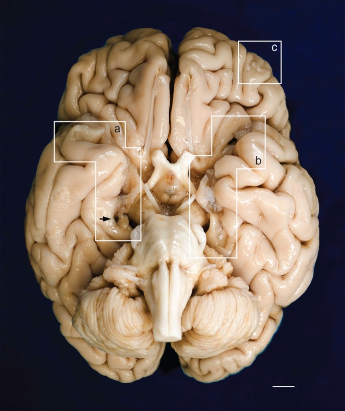
The fixed specimen was photographed after removal of the leptomeninges. Evidence of the surgical lesions in the temporal lobes is highlighted by white geometric contours (a, b). A mark produced by the oxidation of one of the surgical clips inserted by Scoville is visible on the parahippocampal gyrus of the right hemisphere (black arrow). (c) encloses a lesion in the orbitofrontal gyrus that affects the cortex and WM. Marked cerebellar atrophy is consistent with H.M.’s long-term treatment with phenytoin. Scale bar, 1 cm.
The results of our examination are based on 2,401 digital anatomical images and selected corresponding histological sections that were collected at an interval of 70 μm over the course of an uninterrupted 53-hour procedure. The series of digital images of the block’s surface was obtained using a digital camera mounted directly above the microtome stage. Volumetric reconstruction from these images was the basis for subsequent visualization and 3D measurements along arbitrary planes. The dissection of the brain was video-recorded and streamed live on the web to permit scientific scrutiny and to foster public engagement in the study 29 .
3D anatomical measurements in the MTL
Using the 3D measurement tools in AMIRA (FEI Visualization Science Group, Burlington, MA, USA) we calculated the distance in each hemisphere from the anterior tip of the temporal lobe to the posterior boundary of the surgical lesion on each side. This limit was marked by the most anterior coronal anatomical images that did not show any sign of disruption in the normal anatomy, which was confirmed in corresponding stained histological slices. The lesion followed a straight, but slightly oblique path relative to the long axis of each hemisphere; measured along this axis, its length was 54.5 mm and 44.0 mm in the left and right hemispheres, respectively. The value for the left hemisphere was consistent with earlier measurements made on H.M.’s MRI scans acquired in 1992–1993 (ref. 19 ) and 2002–2004 (ref. 20 ). The lesion in the right hemisphere as measured in our postmortem data was 7 mm shorter than in the above-mentioned reports that were based on in vivo imaging.
The borders of the surgical resection were clearly demarcated in the anatomical and histological images; the latter clearly showed that the WM underlying the excised medial temporal cortex was also damaged ( Fig. 2 ). We identified a small portion of the superior-most region of the EC in both hemispheres based on its distinctive cytoarchitecture; specifically, 0.03 cm 3 and 0.11 cm 3 for the left and right hemispheres, respectively (these values correspond approximately to 1.7% and 6.5% of the normal volume, based on published MRI estimates 30 ). Portions of the centromedial nucleus of the amygdala were also preserved ( Fig. 2h,i ).
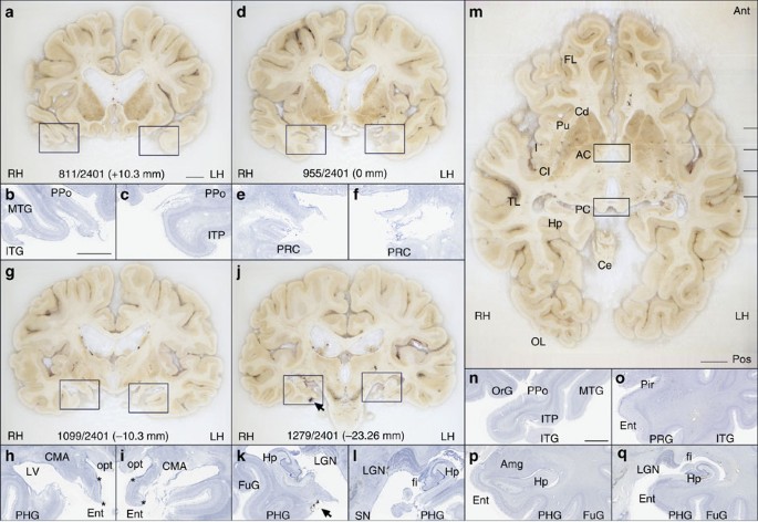
( a , d , g , j ) Cross-sectional anatomy of patient H.M.’s MTL shown at four different levels. The values below the tissue indicate the section number and in parenthesis, the distance from the origin of the standard coordinate system 23 (positive if the level is anterior to the anterior commissure, negative if posterior). Scale bar, 1 cm. ( b , c ), ( e , f ), ( h , i ), ( k , l ): close-up images acquired from thionin-stained tissue slices; these panels illustrate histological detail for the selection boxes in a , d , g , j , respectively. Scale bar, 5 mm. ( m ) Horizontal cross-sectional view reconstructed orthogonally from the original coronal images showing the correct alignment of the anterior and posterior commissures. ( n – q ): normal anatomy and histology of the MTL at the levels shown for the brain of patient H.M. The images were derived from brain slices belonging to a neurologically normal, age-matched individual. Scale bar, 5 mm. PPo, planum polare; MTG, middle temporal gyrus; ITG, inferior temporal gyrus; ITP, inferior temporopolar cortex; PRC, perirhinal cortex; CMA, centromedial amygdala; opt, optic tract; LV, lateral ventricle; PHG, parahippocampal gyrus; Ent, entorhinal cortex; LGN, lateral geniculate nucleus; Hp, hippocampus; FuG, fusiform gyrus; fi, fimbria; SN, substantia nigra; FL, frontal lobe; Cd, caudate; Pu, putamen; I, insula; AC, anterior commissure; Cl, claustrum; TL, temporal lobe; PC, posterior commissure; Ce, cerebellum; OL, occipital lobe; OrG, orbital gyrus; Pir, pirifom cortex; Amg, amygdala. The asterisks delimit the entorhinal cortex.
The extent of the spared hippocampus measured along the horizontal axis of the brain (which in our study coincided with the AC–PC line) was 23.6 mm in the left hemisphere and 24.3 mm in the right. Our measurements included the alveus and the thin band of WM that encapsulates the posterior end of the hippocampus; the fimbria was excluded from our delineations. The discrepancy between these new values and those obtained from earlier MRI data (19 and 22 mm, respectively) is small and can be explained by differences in the quality of the image data 19 . These linear measurements, however, do not fully account for the geometry of H.M.’s spared hippocampus. The high-resolution anatomical volume that we created from microtome images allowed us to inspect the MTL from multiple angles, revealing that the posterior hippocampus was bent steeply in the dorso-medial direction ( Fig. 3 ). This curvature is a normal feature of the periventricular portion of the hippocampus in the human brain, while the most anterior portion, which is immediately posterior to the amygdala and adjacent to the EC, is aligned to the horizontal plane (or major axis) of the hemisphere.
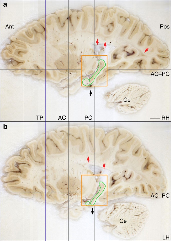
The grey lines intersect at the origin of the origins of the standard coordinate system 23 used for the orientation of the specimen. The blue line indicates the most anterior level of the temporal lobes (the temporal poles, which are damaged are not shown in this image). The orange rectangle represents the bounding box that contains the posterior spared hippocampus (outlined in green), the widest extent of which is at a more medial level than this image shows. The black arrows identify the level at which a surgical clip was positioned on a blood vessel. The red arrows indicate the presence of lesions in the subcortical WM. AC, plane of the anterior commissure; PC, plane of the posterior commissure; AC–PC, ideal plane at the level of both the AC and PC; Ce, cerebellum; LG, lateral geniculate nucleus; Pu, putamen; TP, temporal pole; RH, right hemisphere; LH, left hemisphere. Scale bar, 1 cm.
Measuring the dorso-ventral oblique length of the posterior segment of H.M.’s hippocampus rather than along the AC–PC line produced higher values, specifically, 36.0 mm for the left hemisphere and 40.0 mm for the right. The values were greater when our calculations took curvature into account: in this case, the actual ‘geodesic’ length of the preserved hippocampus amounted to 45.4 mm in the left hemisphere and 47.2 mm in the right ( Fig. 4 ).
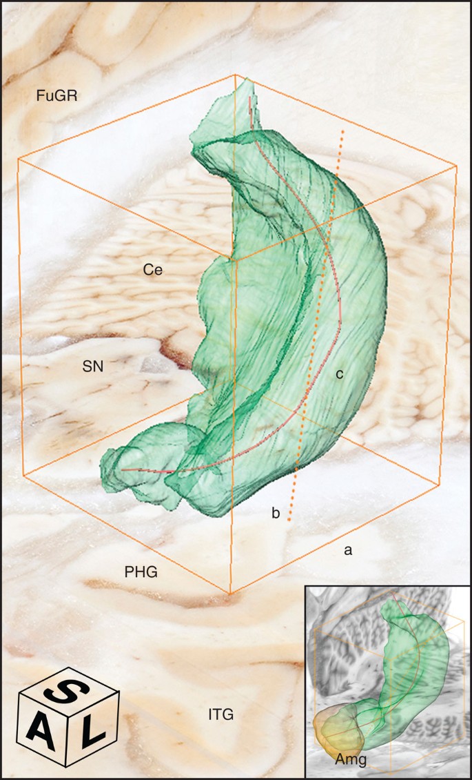
The orange bounding box delimits the dimensions of the structure; different segments indicate different measurements; a: anterior-to-posterior extent in the coronal plane, b (dotted): major diagonal axis, c: anatomical length. The compass cube (A: anterior, L: lateral, S: superior) also functions as scale bar: 5 mm. The insert at the bottom right shows a similarly constructed model from the brain of a neurologically normal subject (78-year-old female donor; volume of the right hippocampus=3.04 mm 3 ; total brain weight 1,278). Ce, cerebellum; FuGR, fusiform gyrus of right hemisphere; IT, inferior temporal gyrus; SN, substantia nigra; Amg, amygdala.
The fact that multiple results can be obtained based on different measuring criteria makes it necessary to establish a clear terminology to describe the anatomy of H.M.’s lesion and the remaining hippocampus in relation to previous reports. In the context of this communication, we refer to the extent of H.M.’s hippocampus and related structures as their linear span in the rostro-caudal direction. This measure can be obtained simply by multiplying the number of tomographic images by the interval between them (that is, slice thickness). The anatomical length of the hippocampus is different and depends on its 3D shape and orientation in the brain. With this distinction in mind, our findings can be more easily reconciled with previous reports that were based on low-resolution scans, where only the extent of the hippocampus was actually measured 19 .
The level of sampling and image quality afforded by the current study represents a significant advance over the first clinical MRI that was performed with H.M. 19 . At that time, the two-dimensional (2D) MRI sequences produced 4–5-mm thick slabs with a 1-mm gap between each slice and an in-plane resolution slightly better than 1 mm per voxel; a T1-weighted 3D MRI scan was also acquired and produced non-isotropic voxels (1 × 1 × 3.2 mm). In these early scans, the boundaries of the posterior hippocampus were blurred by partial volume effects (the presence of multiple tissue types or structures in single large MRI voxels).
In a more recent MRI study with H.M., Salat et al. 20 calculated the volume of tissue ascribed to the posterior hippocampus (voxel size: 1 mm × 1 mm × 1.3 mm) and obtained values of 0.65 cm 3 for the left hemisphere and 0.88 cm 3 for the right. We repeated these measurements based on manual delineations made on three orthogonal views of the digital anatomical reconstruction, and determined that 2.02 cm 3 of hippocampal tissue (including the cornu ammonis, the dentate gyrus and the subiculum) was spared in the left hemisphere and 1.96 cm 3 in the right. The comparison should be interpreted with caution in view of the fact that sufficient normative data on hippocampal volume based on our new postmortem methodology is not yet available. The labelled fields representing the posterior hippocampus in each hemisphere were used to compute triangulated surface models for visualization and shape analyses ( Fig. 4 ).
Regularly spaced tissue sections through the brain were selected at an interval of 1.26 mm; these were mounted on large-format glass slides (5 × 7 in) and stained using thionin. Nissl staining with thionin showed preservation of neuronal cell bodies in CA1, CA2, CA3, CA4 and the subiculum of the hippocampal formation, posterior to the surgical lesion ( Fig. 5 ).
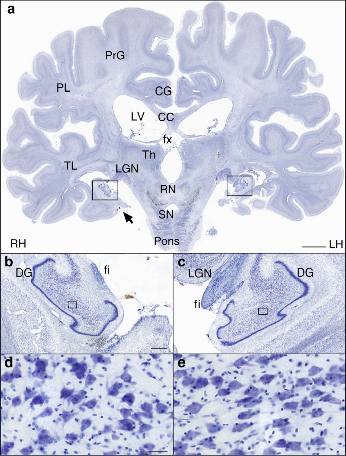
( a ) Whole section. Scale bar, 1 cm. ( b , c ) Higher-magnification cross-sectional image of the dentate gyrus of the spared hippocampus. The fimbria is visible below the lateral geniculate nucleus. Scale bar, 1 mm. ( d , e ) × 20 magnification image of neurons in the CA4 region of the hippocampus (in the location of the box). Scale bar, 50 μm. PrG, precentral gyrus; PL, parietal lobe; CG, cingulate gyrus; LV, lateral ventricle; cc, corpus callosum; fx, fornix; Th, thalamus; TL, temporal lobe; LG, lateral geniculate nucleus; RN, red nucleus; SN, substantia nigra; DG, dentate gyrus; fi, fimbria.
Given that H.M. required surgery because of epilepsy and that there was partial response to the surgical intervention, it is of interest that the typical neuropathologic hallmarks associated with idiopathic temporal lobe epilepsy (granule cell dispersion in the dentate gyrus, neuronal loss from the pyramidal cell layer particularly in area CA4) were not present in the residual hippocampus.
Pathologic anatomy beyond the MTL
While the general size and cortical folding of the cerebral hemispheres appeared normal for an individual of H.M.’s age, multiple WM lesions consistent with lacunar infarctions were present. Cerebellar atrophy was also evident, likely a consequence of long-term exposure to Dilantin (phenytoin sodium), which was part of H.M.’s seizure management pre- and postoperatively 31 , 32 .
Review of the MRI scans and histological sections demonstrated a spectrum of additional lesions in the WM ( Fig. 6 ). We also discovered a small focal lesion in the left lateral orbital gyrus, which was visible on the surface ( Fig. 1 c) and involved both cortex and underlying WM ( Fig. 7 ).
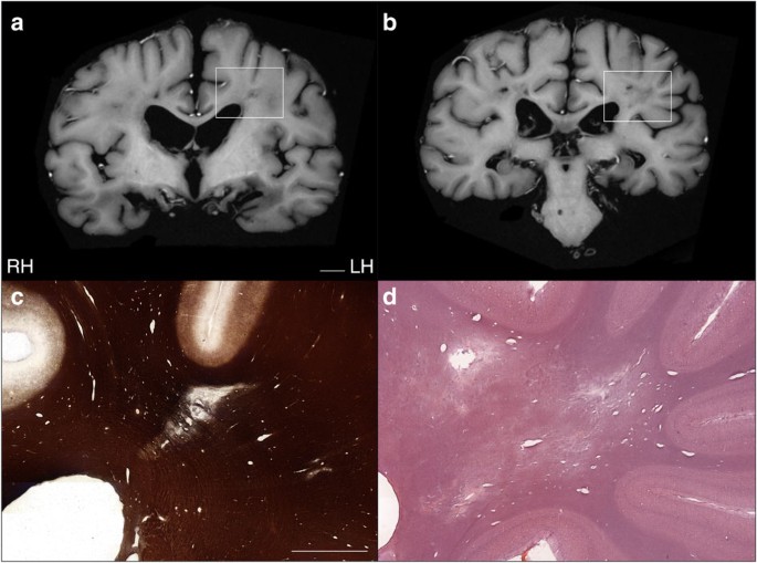
( a , b ) Lesions in the deep WM were visible in postmortem T1-weighted MRI images acquired ex situ (scale bar, 1 cm) and were confirmed by myelin silver-impregnation 40 , 46 ( c ) and haematoxylin and eosin (H&E) staining ( d ). Scale bar, 5 mm.

The reconstructed anatomical volume revealed a small lesion in the lateral orbital gyrus of the left frontal lobe. This is the only lesion affecting the gross structure of the cerebral cortex outside of the MTL. ( a ) T1-weighted MRI of the fixed brain acquired ex situ before cryo-sectioning. Scale bar, 1 cm. The same lesion is shown in histological material that was stained for myelinated fibres 40 , 46 ( b ) and neuronal cell bodies ( c ). Scale bar, 1 mm.
The 3D microscopic model of H.M.’s brain contained clues that help understand the surgery performed in 1953. Scoville approached the MTL of both hemispheres through two small trephine holes ( ∼ 3.8 cm in diameter) drilled above the orbits 17 . The ablation of the tip of the temporal lobe, the uncus and the amygdala was made with a scalpel; the sharp edge of the resections is noticeable in the anatomical and histological images ( Fig. 2d–f ). The more posterior MTL tissue was removed by suction; the fact that the periventricular portion of the hippocampus escaped the ablation can be explained by tracing the trajectory of the suction tube. Based on the reconstructed volume, it is clear that Scoville reached beyond and below the posterior hippocampus as indicated by the position of the surgical clip in the sagittal view of each hemisphere ( Fig. 3 ), although the placement of the clips was also contingent on the pattern of the surface vasculature.
We could reliably identify only a very minimal amount of EC in H.M.’s brain based on cytoarchitecture. The EC (via its superficial layers) is the gateway to the hippocampus for the inflow of information from the cerebral cortex and subcortical nuclei. The hippocampus has reciprocal connections to neocortical areas that deliver the original input via the deep layers of the EC and parahippocampal gyrus, creating a loop that supports the consolidation of long-term memories. Our study confirmed that this circuit was severely compromised by the ablation.
The hippocampus and EC also connect directly (that is, monosynaptically) to specific brain regions such as the nucleus accumbens, amygdala, cingulate cortex and orbitofrontal cortex. Input to and from the hippocampus is arranged along its anatomical length (as defined in this communication) according to an anterior-to-posterior functional gradient. For example, the amygdala is connected to anterior sections of the EC and hippocampus, while the visual cortex (largely residing in the occipital, parietal, lateral and ventral temporal lobes) is functionally linked to the posterior hippocampus. This organization is also reflected in the topography of differential gene expression and specific functional activation 33 , 34 , 35 . According to these maps, and based on the gradient of connectivity within the MTL and with other brain structures, the hippocampus appears broadly subdivided into an anterior segment that supports emotion, stress and sensorimotor integration, and a posterior part dedicated to declarative memory and neocortex-supported cognition 36 , 37 . According to this model, the almost complete removal of the EC in both hemispheres, resulting in severe disconnection of the remaining hippocampus, would have made a more significant contribution to H.M.’s declarative memory impairment than the ablation of the anterior hippocampus.
The excision of the anterior hippocampus, together with the bulk of the amygdala, may explain H.M.’s dampened expression of emotions, poor motivation and lack of initiative 19 . The fact that he was impaired in reporting internal states such as pain, hunger and thirst and his apparent lack of initiative was ascribed to the almost complete removal of the amygdala, although it remains difficult to distinguish between direct injury to the amygdala and disruption of connections into and out of this complex structure 6 , 38 . The sparing of the posterior segment of the parahippocampal cortex may validate observations of H.M.’s preserved visual perception, perceptual learning and perceptual priming 6 , 39 . A detailed architectonic survey of occipital, parietal and ventral temporal areas 40 , 41 could provide new clues about the neural circuits H.M. engaged during the performance of numerous perceptual tasks.
Histological staining revealed neuronal integrity in the CA4 field of the remaining hippocampus 42 and a normal compact pattern of the granule cell layer of the dentate gyrus ( Fig. 5 ). These findings raise interesting questions regarding the functional viability of H.M.’s remaining hippocampal tissue because according to established models of hippocampal connectivity, the gap in the MTL created by the surgery would have interrupted crucial connections to and from the posterior hippocampus. Additional quantitative analyses involving unbiased stereological methods can provide information regarding neuronal size and number 43 in this region. Combined with molecular approaches such as antibody staining to assess synaptic density, patterns of gliosis, and the integrity of afferent and efferent fibres, these studies could contribute further insight on the intrinsic and long-range connectivity of the human hippocampus.
The lesion on the left frontal lobe was circumscribed to a small area within the left lateral orbital gyrus ( Fig. 1 c), and it involved the cortical ribbon as well as the underlying WM ( Fig. 7 ). Because of the removal of all leptomeninges as part of the preparation of the brain prior to sectioning, it is not possible to determine the status of the immediate subpial zone in the area of the lesion, which would have assisted in discriminating a mechanical laceration from a vascular injury. As mentioned above, Scoville performed the removal of deep MTL structures while each frontal lobe was retracted to provide access to the deeper temporal structures; thus, judging from the orientation of the ablation with respect to the position of the trephine holes, the scar in the left prefrontal cortex could plausibly have been created at the time of the surgery.
In the course of the initial fixation, when the specimen was suspended upside down, the damaged portion of the temporal lobes collapsed; this geometrical artefact of fixation, combined with the relative shrinkage of the whole specimen during histological processing, should be considered when our resultsare compared with previous MRI scans obtained in vivo . It should be noted, however, that fixation, combined with cryoprotection, produced only an estimated 5–7% reduction in volume of the whole brain and these gross changes did not affect the morphology of the spared hippocampus.
Attempts to provide structure–function correlations based on the postmortem evaluation of H.M.’s MTL require a parallel assessment of age-related neuropathologic processes that developed progressively in his brain. MRI demonstrated extensive WM T2-hyperintensities, which were not related to the surgery; these were confirmed in histological sections ( Fig. 6 ). Although H.M. suffered a minor head injury before age 10, this kind of event does not typically produce long-lasting traumatic injuries that can be subsequently detected radiologically, especially in the absence of clinical signs. Rather, the nature and distribution of observed WM pathology suggest the manifestation of microvascular disease associated with hypertension. Diagnostic neuropathologic evaluation is still required to determine the types and burden of abnormalities and disease processes that occurred independently of the surgery as a result of aging and related to other aetiologies.
The purity and severity of H.M.’s memory impairment, combined with his willingness to participate in testing, made his case study uniquely influential in the field of memory research, as reflected in the broad range of scientific publications built on the studies to which he contributed 19 . During life, H.M. was the best-known and possibly the most studied patient in modern neuroscience; the availability of a large and organized collection of slices and digital anatomical images through the whole brain provides an unprecedented opportunity for collaborative and retrospective studies to continue 22 , 44 . The archive of images will also constitute a permanent digital trace for the case that defined two generations of memory research and that will likely remain as emblematic for neuroscience in the future as it is today.
Ethics statement
The study was reviewed and approved by one of UC San Diego’s Institutional Review Boards in accordance with the requirements of the Code of Federal Regulations for the Protections of Human Subjects. The removal and preservation of H.M.’s brain at autopsy, as well as disclosure of protected health information about him, was authorized by his legally appointed representative.
Preparation of the brain
Shortly after his death, H.M’s body was transported to the Anthinoula A. Martinos Center for Biological Imaging, in Charlestown, MA, where 9 h of in situ scanning was performed 21 . Following this imaging session, an autopsy was conducted; the brain removed, weighed (the total brain weight was 1,300 g) and then fixed in standard buffered formalin (4% formaldehyde; postmortem interval of ∼ 14 h). The brain was fixed for 10 weeks at 4 °C with three changes of fixative during that time; it was suspended upside down, hung by the basilar artery. When the tissue was firm enough, the brain was immersed in fixative laying on a cushion of hydrophilic cotton. Subsequently, multiple series of MRI scans of the fixed specimen were acquired in 3T and 7T scanners 21 .
H.M.’s brain was transported to The Brain Observatory in February 2009, where fixation in formaldehyde continued for 2 more months. After this additional period of fixation, the brain was imaged in a 1.5 T General Electric (GE) Excite MRI scanner using an eight-channel transmit-receive head coil ( Figs 6a,b and 7a ). We used a T1-weighted, 3-D Inversion-Recovery Fast Gradient Echo (IR-FSPRG) sequence (image matrix: 512 × 512; Slice thickness: 1mm; FOV: 256; TE: 15; 18 excitations, or NEX. Duration of the scan: ∼ 7 hours). During the scan the brain was soaked in formaldehyde solution and contained in a watertight Plexigas chamber designed to fit inside the coil above mentioned. Following MRI, the brain was immersed in solutions of increasing concentration of sucrose; specifically, 10, 20 and 30% in 4% formaldehyde solution, phosphate-buffered to pH 7.4. Treatment with sucrose lasted ∼ 10 months: it was meant to minimize the formation of ice crystals and reduced the risk of creating cracks when the brain was frozen.
Following fixation and cryoprotection, the leptomeninges were removed and the brain was embedded in a rectangular cast of 10% gelatin. The whole-brain-gelatin block was chilled to −40 °C by immersion in recirculating isopentane (2-methylbutane) using a custom-built insulated chamber. As the brain froze, the temperature of the solvent was constantly monitored and recorded via a probe connected to a personal computer running a custom programme (Labview, National Instruments Inc. Austin, TX, USA). The brain was thoroughly frozen after ∼ 5 h, when the temperature of the block and that of the surrounding fluid reached a stable equilibrium, based on probe data. The frozen block was firmly attached to the stage of a heavy-duty sliding microtome (Polycut SM 2500; Leica Microsystems Inc., Buffalo Grove, IL) and kept frozen by an assembly of interlocking hollow cuffs that transferred heat through a continuous flow of cold ethanol; the alcohol was recirculated and maintained at −40 °C by a battery of industrial chillers.
A second apparatus was mounted to the microtome to support a digital camera and lighting system directly above the microtome stage; it was designed to acquire high-resolution images of the surface of the block (gelatin and brain) through the entire slicing procedure. These images were the basis for 3D reconstruction and currently serve as a digital catalogue representing the collection of tissue slices.
Image acquisition and volume reconstruction
Tomographic images were acquired using a digital single-lens reflex camera (Nikon D700, Nikon Inc., Melville, NY) bearing a 35-mm lens (AF NIKKOR 35 mm f/2D), which was connected to a PC workstation and controlled remotely. A pair of opposite, light-emitting diode arrays were pointed at the surface of the block at an angle of 45°, providing constant illumination during the cutting procedure. The image acquisition sequence was fully automated; exposure was triggered before each new stroke was initiated. Image files were saved directly on disk in raw (uncompressed) format measuring 4,256 × 2,832 pixels (equivalent to a resolution of 46 μm per pixel). The microtome camera assembly was designed to trigger each exposure every time the microtome stage stopped at a fixed position so that the resulting image stack was registered mechanically. Dedicated team members maintained a log detailing file acquisition and inspected every newly acquired image in real time. Later, we used the quality control report to identify image artifacts in the series so that ad hoc post-processing routines for 3D reconstruction could be applied.
A Canon XLH1A digital HD camcorder (Canon U.S.A., Inc. Lake Success, NY) was used to record the procedure and provide a live feed of a close up of the brain within the microtome assembly on the web. Two additional views, of the microtome console and a wide-angle view of the lab, were provided via Microsoft LifeCam Cinema webcams (Microsoft Inc. Redmond, WA). The video from each of the three views was converted to flash with Adobe Flash Media Live Encoder 3.1 and streamed using Adobe Flash Media Server 3.5 (Adobe Systems Incorporated, San Jose, CA, USA).
Images acquired during the cutting procedure were transferred to a Dell PowerEdge 7500 workstation running a Windows 7, 64-bit operating system with 192 GB of DDR3 dynamic random-access memory (DRAM). The digital 3D volume representing the whole brain was assembled using AMIRA. Scaling factors were derived from the in-plane resolution of the images (converted from pixels to mm) and the slicing interval (that is, section thickness). The resolution of the data set was 46 × 46 × 70μm and occupied ∼ 56 GB on disk. Access to large amounts of DRAM allowed for the efficient manipulation and inspection of the entire anatomical volume. Measurements were conducted on a volume created from images that were down-sampled to 2,550 × 2,550 pixels (voxel dimensions were x : 56.9 μm; y : 56.9 μm and z : 70 μm).
The images were cropped to the edge of the gelatin block and corrected for any changes in brightness produced by fluctuations in ambient lighting that occurred during the 3-day procedure. As the ratio between brain tissue and gelatin matrix varied considerably at different levels of the series of anatomical images, we created a mask for brain tissue only and used it to adjust levels across the series. This process produced a uniform volumetric data set that was consistent in terms of luminosity and contrast in any arbitrary plane. Close inspection of the final 3D reconstruction identified occasional shifts in alignment, most likely due to accidental mechanical shifts in the equipment. Consequently, the position of outlier images was corrected by applying 2D rigid transformations (translation and rotation). The transformation matrix was computed with a local search algorithm that used a least-squares metric to compare intensities between each pair of consecutive images.
3D measurements of the hippocampus
The delineation of hippocampal structures was performed on the 3D reconstruction created from the series of digital images acquired in the course of the slicing procedure. Three main orthogonal planes were visualized concurrently in the segmentation editor of AMIRA to ensure accuracy. In the coronal plane, the hippocampus was identified starting at the posterior-most level of the mammillary bodies and the structure withered at the level of the inferior colliculi. The delineation of the inferior medial border (that is, the border between the subiculum and parahippocampal cortex) was aided by the identification of the perforant pathway in the anatomical images and changes in cytoarchitecture in corresponding Nissl-stained sections. Sagittal views afforded a clear demarcation of the hippocampus’ posterior border ( Fig. 3 ); the alveus was included in the calculation of the volume 45 . The volume of the hippocampus in each hemisphere was calculated as the number of tissue voxels comprised in the delineations multiplied by the individual voxel dimensions in mm.
The anatomical central axis of the hippocampus was defined as a 3D curve fit with a given stiffness along a path through the field of labelled voxels. We calculated a straight-line path using principal component analysis of a binary segmentation of the region of interest. The 3D line was sampled at uniform intervals (10 voxels) to define control points. The first and last control points of the line were set to the 3D locations defined by the furthermost extent of the object along the line. To improve the location of the control points, we sectioned the object at positions halfway between each pair of control points, using a plane that was orthogonal to the curve’s tangent direction at that location, and yielding one section for each control point. For each section of the binary object, we calculated its centre of mass and moved the corresponding control point towards that location. After the adjustments of all control points, a curve-smoothing operation was executed that used the predefined stiffness constraint on the curve. The extent of smoothing controlled the stiffness attached to the curve. The sectioning, adjustment and smoothing steps were repeated until convergence was achieved. This expectation–maximization approach iteratively adjusted the location of the curve’s control points towards a solution that intersected the object uniformly and had a given stiffness. The resulting 3D curve defined the ‘geodesic’ axis of the region of interest, and it was the basis for the actual anatomical length measurement.
Authors contributions
J.A. designed the neuroanatomical study and major instrumentation. M.P.F. conducted the brain extraction at autopsy assisted by J.A. Serial sectioning and tomographic imaging of the brain specimen was performed by J.A. with the assistance of N.M.S., P.M., N.T., J.K., A.G. and N.B., H.B., N.M.S., P.M. and J.A. developed the 3D model of the brain from cross-sectional image data acquired during the dissection. H.B. developed the 3D measurement and image registration tools. J.A., P.M. and C.S produced the stained histological slides. J.A. wrote the manuscript with significant editing contributions by S.C. and M.P.F. Photographs and figures were created by J.A. and C.S. Additional background research was conducted by R.K.
Additional information
How to cite this article: Annese, J. et al. Postmortem examination of patient H.M.’s brain based on histological sectioning and digital 3D reconstruction. Nat. Commun. 5:3122 doi: 10.1038/ncomms4122 (2014).
Scoville, W. B. & Milner, B. Loss of recent memory after bilateral hippocampal lesions. J. Neurol. Neurosurg. Psychiatr. 296 , 1–22 (1957).
Google Scholar
Corkin, S. Lasting consequences of bilateral medial temporal lobectomy: clinical course and experimental findings in H.M. Sem. Neurol. 4 , 249–259 (1984).
Article Google Scholar
Corkin, S. Tactually-guided maze learning in man: Effects of unilateral cortical excisions and bilateral hippocampal lesions. Neuropsychologia 3 , 339–351 (1965).
Corkin, S. Acquisition of motor skill after bilateral medial temporal-lobe excision. Neuropsychologia 6 , 255–265 (1968).
Woodruff-Pak, D. S. Delay and trace paradigms. Behav. Neurosci. 107 , 911–925 (1993).
Article CAS Google Scholar
Milner, B., Corkin, S. & Teuber, H.-L. Further analysis of the hippocampal amnesiac syndrome: 14-year follow-up study of H.M. Neuropsychologia 6 , 215–234 (1968).
Keane, M. M., Gabrieli, J. D. E., Mapstone, H. C., Johnson, K. A. & Corkin, S. Double dissociation of memory capacities after bilateral occipital-lobe or medial temporal-lobe lesions. Brain 118 , 1129–1148 (1995).
Milner, B. Memory and the medial temporal regions of the brain. In Biology of Memory (eds Pribram K. H., Broadbent D. E. )29–50Academic Press (1970).
Milner, B. The medial temporal lobe amnesic syndrome. Psychiatr. Clin. N. Am. 28 , 599–611 (2005).
Cohen, N. J. & Squire, L. R. Preserved learning and retention of pattern analyzing skill in amnesia: dissociation of knowing how and knowing that. Science 210 , 207–209 (1980).
Article CAS ADS Google Scholar
Steinvorth, S., Levine, B. & Corkin, S. Medial temporal lobe structures are needed to re-experience remote autobiographical memories: evidence from H.M. and W.R. Neuropsychologia 43 , 479–496 (2005).
Squire, L. R. & Wixted, J. T. The cognitive neuroscience of human memory since H.M. Ann. Rev. Neurosci. 34 , 259–288 (2011).
Tulving, E. Episodic and semantic memory. In: Organization of Memory (eds Tulving E., Donaldson W. )381–402Academic Press (1972).
Gabrieli, J. D. E., Cohen, N. J. & Corkin, S. The impaired learning of semantic knowledge following bilateral medial temporal-lobe resection. Brain Cogn. 7 , 157–177 (1988).
O’Kane, G., Kensinger, E. A. & Corkin, S. Evidence for semantic learning in profound amnesia: an investigation with patient H.M. Hippocampus 14 , 417–425 (2004).
Scoville, W. B. The limbic lobe in man. J. Neurosurg. 11 , 64–66 (1954).
Scoville, W. B., Dunsmore, R. H., Liberson, W. T., Henry, C. E. & Pepe, A. Observations on medial temporal lobotomy and uncotomy in the treatment of psychotic states. Res. Pub. Assoc. Res. Nerv. Ment. Dis. 31 , 347–373 (1953).
CAS Google Scholar
Scoville, W. B. Amnesia after bilateral mesial temporal-lobe excision: Introduction to case H.M. Neuropsychologia 6 , 211–213 (1968).
Corkin, S., Amaral, D. G., González, R., Johnson, K. & Hyman, B. T. H.M.’s medial temporal lobe lesion: findings from magnetic resonance imaging. J. Neurosci. 17 , 3964–3979 (1997).
Salat, D. H. et al. Neuroimaging H.M.: a 10-year follow-up examination. Hippocampus 16 , 936–945 (2006).
van der Kouwe, A. et al. Technical description of post-mortem magnetic resonance neuroimaging of H.M. Program No. 714.17. 2010 Neuroscience Meeting Planner. San Diego, CA: Society for Neuroscience, 2010. Online.
Annese, J. Deconstructing Henry: the neuroanatomy and neuroinformatics of the brain of the amnesic patient H.M. Program No. 397.18. 2010 Neuroscience Meeting Planner. San Diego, CA: Society for Neuroscience, 2010. Online.
Talairach, J. & Tournoux, P. Co-Planar Stereotaxic Atlas of the Human Brain Thieme Medical Publishers (1988).
Wilson, B. A., Baddeley, A. D. & Kapur, N. Dense amnesia in a professional musician following herpes simplex virus encephalitis. J. Clin. Exp. Neuropsychol. 17 , 668–681 (1995).
Stefanacci, L., Buffalo, E. A., Schmolck, H. & Squire, L. R. Profound amnesia after damage to the medial temporal lobe: a neuroanatomical and neuropsychological profile of patient E.P. J. Neurosci. 20 , 7024–7036 (2000).
Insausti, R., Annese, J., Amaral, D. G. & Squire, L. R. Human amnesia and the medial temporal lobe illuminated by neuropsychological and neurohistological findings for patient E.P. Proc. Natl Acad. Sci. USA 110 , E1953–E1962 (2013).
Damasio, A. R., Eslinger, P., Damasio, H., van Hoesen, G. W. & Cornell, S. Multimodal amnesic syndrome following bilateral and basal forebrain damage. Arch. Neurol. 42 , 252–259 (1985).
Annese, J. The importance of combining MRI and large-scale digital histology in neuroimaging studies of brain connectivity and disease. Front. Neuroinform. 6 , 1–5 (2012).
Buchen, L. Famous brain set to go under the knife. Nature 462 , 403 (2009).
Insausti, R. et al. MR volumetric analysis of the human entorhinal, perirhinal, and temporopolar cortices. Am. J. Neuroradiol. 19 , 659–671 (1998).
CAS PubMed Google Scholar
De Marcos, F. A., Ghizoni, E., Kobayashi, E., Li, L. M. & Cendes, F. Cerebellar volume and long-term use of phenytoin. Seizure 12 , 312–315 (2003).
Ghatak, N. R., Santoso, R. A. & McKinney, W. M. Cerebellar degeneration following long-term phenytoin therapy. Neurology 26 , 818–820 (1976).
Zhao, X. et al. Transcriptional profiling reveals strict boundaries between hippocampal subregions. J. Comp. Neurol. 441 , 187–196 (2001).
Thompson, C. et al. Genomic anatomy of the hippocampus. Neuron 60 , 1010–1021 (2008).
Fanselow, M. & Dong, H. Are the dorsal and ventral hippocampus functionally distinct structures? Neuron 65 , 7–19 (2010).
Small, S., Nava, A., DeLaPaz, R., Mayeux, R. & Stern, Y. Circuit mechanisms underlying memory encoding and retrieval in the long axis of the hippocampal formation. Nat. Neurosci. 4 , 442–449 (2001).
Lavenex, P. & Amaral, D. Hippocampal-neocortical interaction: a hierarchy of associativity. Hippocampus 10 , 420–430 (2000).
Hebben, N., Corkin, S., Eichenbaum, H. & Shedlack, K. Diminished ability to interpret and report internal states after bilateral medial temporal resection: case H.M. Behav. Neurosci. 99 , 1031–1039 (1985).
Corkin, S. What’s new with the amnesic patient H.M.? Nat. Rev. Neurosci. 3 , 153–160 (2002).
Annese, J., Pitiot, A., Dinov, I. & Toga, A. A myeloarchitectonic method for the structural classification of cortical areas. NeuroImage 21 , 15–26 (2004).
Annese, J., Gazzaniga, M. & Toga, A. Localization of the human cortical visual area MT based on computer-aided histological analysis. J. Neurosci. 15 , 1044–1053 (2005).
Duvernoy, H. M. The Human Hippocampus: Functional Anatomy, Vascularization and Serial Sections with MRI, Third Edn Springer (2005).
West, M. J. & Gundersen, H. J. Unbiased stereological estimation of the number of neurons in the human hippocampus. J. Comp. Neurol. 296 , 1–22 (1990).
Annese, J. From the jar to the world wide web: designing a public digital library for the human brain. Interdiscipl. Sci. Rev. 38 , 222–231 (2013).
Insausti, R. & Amaral, D. G. Hippocampal formation. In The Human Nervous System 2nd edn (eds Paxinos G., Mai J. K. )871–914Academic Press (2004).
Gallyas, F. Silver staining of myelin by means of physical development. Neurol. Res. 1 , 203–209 (1979).
Download references
Acknowledgements
The work was supported by grants from the National Science Foundation (NSF—SGER 0714660, J.A., Principal Investigator), the Dana Foundation (Brain and Immuno-Imaging Award, 2007-4234, J.A., Principal Investigator) and by private contributions from viewers of the web broadcast of the dissection. In the course of the study, Dr. Annese was in part supported by two research grants from the National Eye Institute, R01 EY018359–02 and ARRA R01 EY018359–02S1 (J.A., Principal Investigator) and the National Institute of Mental Health, R01MH084756 (J.A., Principal Investigator). The authors would like to thank Herbert and Sharon Lurie, M.S., for their generous contribution towards publishing the article on an open access basis. We would like to dedicate the project and this publication to H.M. He transformed his difficult experience into a lifelong contribution to scientific research on the mechanisms supporting human memory. This study also honours all the researchers who conducted studies with H.M. when he was alive, and Brenda Milner, in particular, who first demonstrated the importance of his case. The authors would like to acknowledge the critical contribution of David Malmberg in the development of the instrumentation and thank Dr. Elizabeth Murphy for editorial assistance.
Author information
Natasha Thomas, Junya Kayano and Alexander Ghatan: These authors contributed equally to this work
Authors and Affiliations
The Brain Observatory, San Diego, 92101, California, USA
Jacopo Annese, Natalie M. Schenker-Ahmed, Hauke Bartsch, Paul Maechler, Colleen Sheh, Natasha Thomas, Junya Kayano, Alexander Ghatan, Noah Bresler & Ruth Klaming
Department of Radiology, University of California San Diego, San Diego, 92093, California, USA
Jacopo Annese, Natalie M. Schenker-Ahmed, Hauke Bartsch, Paul Maechler, Colleen Sheh & Ruth Klaming
C.S. Kubik Laboratory for Neuropathology, Massachusetts General Hospital and Harvard Medical School, 02114, Massachusetts, USA
Matthew P. Frosch
Department of Brain and Cognitive Sciences, Massachusetts Institute of Technology, 02139, Massachusetts, USA
Suzanne Corkin
You can also search for this author in PubMed Google Scholar
Corresponding author
Correspondence to Jacopo Annese .
Ethics declarations
Competing interests.
The authors declare no competing financial interests.
Rights and permissions
This work is licensed under a Creative Commons Attribution-NonCommercial-NoDerivs 3.0 Unported License. To view a copy of this license, visit http://creativecommons.org/licenses/by-nc-nd/3.0/
Reprints and permissions
About this article
Cite this article.
Annese, J., Schenker-Ahmed, N., Bartsch, H. et al. Postmortem examination of patient H.M.’s brain based on histological sectioning and digital 3D reconstruction. Nat Commun 5 , 3122 (2014). https://doi.org/10.1038/ncomms4122
Download citation
Received : 13 October 2013
Accepted : 16 December 2013
Published : 28 January 2014
DOI : https://doi.org/10.1038/ncomms4122
Share this article
Anyone you share the following link with will be able to read this content:
Sorry, a shareable link is not currently available for this article.
Provided by the Springer Nature SharedIt content-sharing initiative
This article is cited by
Immunohistochemical field parcellation of the human hippocampus along its antero-posterior axis.
- Emilio González-Arnay
- Isabel Pérez-Santos
- Carmen Cavada
Brain Structure and Function (2024)
High Pressure Hosing-Drone Dynamics and Controls
- Blake Hament
Journal of Intelligent & Robotic Systems (2023)
Convolutional neuronal networks combined with X-ray phase-contrast imaging for a fast and observer-independent discrimination of cartilage and liver diseases stages
- Johannes Stroebel
- Annie Horng
Scientific Reports (2020)
Pavlovian patterns in the amygdala
- Bruno B. Averbeck
Nature Neuroscience (2019)
Dissecting the pathobiology of altered MRI signal in amyotrophic lateral sclerosis: A post mortem whole brain sampling strategy for the integration of ultra-high-field MRI and quantitative neuropathology
- Menuka Pallebage-Gamarallage
- Sean Foxley
- Olaf Ansorge
BMC Neuroscience (2018)
By submitting a comment you agree to abide by our Terms and Community Guidelines . If you find something abusive or that does not comply with our terms or guidelines please flag it as inappropriate.
Quick links
- Explore articles by subject
- Guide to authors
- Editorial policies
Sign up for the Nature Briefing newsletter — what matters in science, free to your inbox daily.
Final dates! Join the tutor2u subject teams in London for a day of exam technique and revision at the cinema. Learn more →
Reference Library
Collections
- See what's new
- All Resources
- Student Resources
- Assessment Resources
- Teaching Resources
- CPD Courses
- Livestreams
Study notes, videos, interactive activities and more!
Psychology news, insights and enrichment
Currated collections of free resources
Browse resources by topic
- All Psychology Resources
Resource Selections
Currated lists of resources
Study Notes
Scoville and Milner (1957)
Last updated 22 Mar 2021
- Share on Facebook
- Share on Twitter
- Share by Email
Effect of hippocampal damage on memory.
Background information: Scoville performed experimental surgery on H.M.’s brain to stop the severe epileptic seizures he had been suffering since a fall off his bicycle many years previously. Specifically, he removed parts of HM's temporal lobes (part of his hippocampus along with it). The seizures reduced drastically but H.M. suffered from amnesia for the rest of his life. Milner, who was a PhD student of Scoville’s, followed up the surgery with cognitive testing for fifty years after the original operation. Hers is a cognitive longitudinal case study of H.M.’s anterograde (after the surgery) and partial retrograde (before the surgery) amnesia. The biological part of the H.M. study is the correlation between the brain damage and the amnesia, which was assumed in the 1950s, and not verified until later brain scans in the 1990s (see Corkin, 1997)
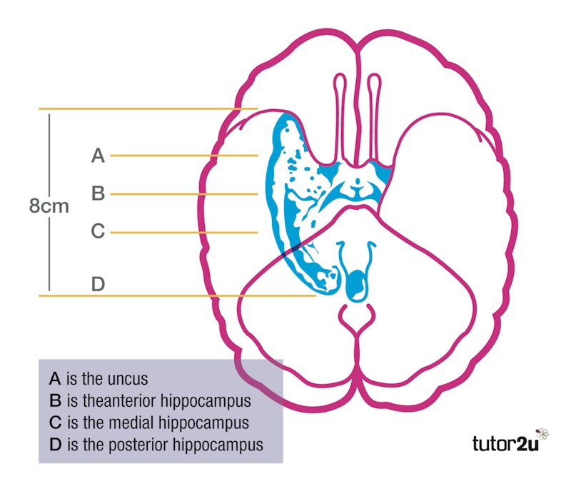
Aim: In 1953 Scoville performed surgery on the then 27-year-old H.M. to cure him of his epileptic seizures. [Note: this is a surgical procedure – it only became a study later when the memory damage was noted].
Method: The surgery involved what was called a partial medial temporal lobe resection. Scoville removed 8 cm of brain tissue from the anterior two thirds of the hippocampus, and believed he “probably destroyed …. the uncus and amygdala” as well (Scoville and Milner, 1957). Once the extent of the memory loss was realised, Scoville and Milner wrote about this, along with the results from this type of surgery on nine other patients, in a prominent neurosurgical journal, and Milner started her cognitive studying of H.M.
Results: H.M. lost the ability to form new memories. This is called anterograde amnesia. He could do a task, and even comment that it seemed easier than he expected, without realising that he had done it hundreds of times before. His anterograde procedural memory was totally affected. He also lost his memory for events that had happened after his surgery: he could not remember moving house, nor that he had eaten a meal thirty minutes previously. He had also suffered some retrograde amnesia of events preceding the surgery, such as the death of his uncle three years before. However, his early childhood memories remained intact. His intelligence also remained as before, at slightly above average.
Conclusion: The surgery to remove part of the hippocampus, the uncus and the amygdala resulted in total anterograde amnesia and partial retrograde amnesia.
Evaluation:
This is the assumption, based on the results with other patients as well as H.M. In the absence at that time of brain-scanning equipment, other possibilities were also present. The high doses of anti-epileptic drug he was taking before, and the lower doses after the surgery, may have resulted in some memory loss. Also, so far as we can see, no memory tests were conducted on H.M. before the surgery, and the initial memory loss was largely reported by his mother, with whom he lived.
For More Study Notes…
To keep up-to-date with the tutor2u Psychology team, follow us on Twitter @tutor2uPsych , Facebook AQA / OCR / Edexcel / Student or subscribe to the Psychology Daily Digest and get new content delivered to your inbox!
- Hippocampus
You might also like
Corkin (1997), fink et al. (1996), maguire et al. (2000), ib psychology (bloa): animal research may inform our understanding of human behaviour, ib psychology (bloa): cognitions, emotions & behaviours are products of the anatomy and physiology of our nervous and endocrine system, our subjects.
- › Criminology
- › Economics
- › Geography
- › Health & Social Care
- › Psychology
- › Sociology
- › Teaching & learning resources
- › Student revision workshops
- › Online student courses
- › CPD for teachers
- › Livestreams
- › Teaching jobs
Boston House, 214 High Street, Boston Spa, West Yorkshire, LS23 6AD Tel: 01937 848885
- › Contact us
- › Terms of use
- › Privacy & cookies
© 2002-2024 Tutor2u Limited. Company Reg no: 04489574. VAT reg no 816865400.
- Skip to main content
- Keyboard shortcuts for audio player
Research News
H.m.'s brain and the history of memory.
Brian Newhouse
In 1953, radical brain surgery was used on a patient with severe epilepsy. The operation on "H.M." worked, but left him with almost no long-term memory. H.M. is now in his 80s. His case has helped scientists understand much more about the brain.
Web Resources
SCOTT SIMON, host:
Fifty years ago this month, the Journal of Neurology, Neurosurgery and Psychiatry published the findings of a remarkable case. A young man who had undergone an experimental brain operation had lost his ability to retain new memories. He could remember things from his life before the operation but any new face or fact, he completely forgot within minutes. Researchers at that time studied him. And it turns out their discoveries opened the modern era of memory research, what's involved every time we say I remember. That young man is now in his 80s. And as Brian Newhouse reports, scientists are still learning from him.
(Soundbite of crickets)
BRIAN NEWHOUSE: If you close your eyes and just listen for a moment, you may find yourself going somewhere, back in memory. Back maybe to a farm or a park or a lake. Other sounds may make those memories sharpen or change. Add still more and you may start to see particular faces or even smell wood smoke. Remember?
(Soundbite of laughter)
NEWHOUSE: This is the power of memory, the system that captures pictures, smells, sounds, events, directions - endless amounts of information every day and then seconds or decades later calls it up for us. Memories - what we've learned and what we've done - in a large sense make us who we are. To appreciate this, think if your ability to form any new memories were suddenly cut off.
Who would you be? By studying people who've lost their memories, scientists have learned enormous amounts about how learning and memory work in healthy brains. And what they used to think was relatively straightforward they've since found it's fascinatingly complex, thanks in large part to one man.
He's the most famous patient in the study of the human brain today. He's written up in textbooks and dozens of scientific papers. The modern era of memory research essentially began with him, yet very few people know his name or have ever seen him, despite the efforts of journalists, filmmakers and TV networks, all of whom have asked to photograph, film or interview him. Outside the circle of his family and caregivers, he's known only by his initials, H.M. His guardians recently agreed to release audio recordings made of him in the early 1990s talking to scientists. This is the first time a wide audience has been able to hear his voice.
Dr. BRENDA MILNER (McGill University) : When you're not at MIT, what do you do during a typical day?
H.M. (Patient): See, that's what I don't - I don't remember things.
Dr. MILNER: Uh-huh.
H.M. was a very pleasant normal young man, but he had suffered from very severe epilepsy all his life, really. It made him unable to hold down his job as an assembly worker. It made him very late in finishing high school, although he was quite intelligent.
NEWHOUSE: Brenda Milner is a British-born neuroscientist at McGill University in Montreal who first met H.M. in the mid-1950s.
Dr. MILNER: He had huge major and minor seizures, you know, huge convulsions, and also many, many lapses of consciousness every few minutes. He was in a very, very hopeless condition with his epilepsy.
NEWHOUSE: Dr. Milner came to know H.M. after Connecticut neurosurgeon William Beecher Scoville performed an experimental operation to help relieve H.M.'s seizures. Dr. Scoville thought if he could remove the part of H.M.'s brain where the seizures originated, it might stop them.
Dr. MILNER: And this operation was carried out when - in '53, 1953, September - when H.M. was 27. The operation did have an enormously beneficial effect on the epilepsy so that H.M. has maybe now one big seizure a year, and so the clinical hunch about the epilepsy was justified. But at obviously a horrendous price.
(Soundbite of piano)
Dr. MILNER: Who is the president of the United States now?
H.M.: That I don't - I couldn't tell you. I don't remember exactly at all.
Dr. MILNER: Is it a man or a woman?
H.M.: I think it's a man.
Dr. MILNER: His initials are G.B. Does that help?
H.M.: No, it doesn't help.
NEWHOUSE: The horrendous price that H.M. paid was a severe case of amnesia. Not the amnesia of Hollywood, where a person forgets everything about his past, but in H.M. it's his ability to acquire new memories, to commit to memory even the simplest events of his day or the world around him, and then to effectively retrieve those memories. Put a finger above your ear. If you were able to push that finger into your head about two inches, you'd be in the area called the medial temporal lobe. There's one on each side of the brain. In the 1950s, Dr. Scoville theorized that these were the general areas involved in H.M.'s epilepsy. But in trying to alleviate H.M.'s seizures, Dr. Scoville removed most of the medial temporal lobes, including much of the hippocampus. This unintentional experiment showed that the hippocampus and medial temporal lobes are where the brain converts short term memory into long term memory.
Dr. MILNER: Do you know what you did yesterday?
H.M.: No, I don't.
Dr. MILNER: How about this morning?
H.M.: I don't even remember that.
Dr. MILNER: Could you tell me what you had for lunch today?
H.M.: I don't know, to tell you the truth.
NEWHOUSE: H.M.'s condition has also helped scientists understand how and where the brain processes different types of memory. Scientists now know that some brain structures are involved in things like phone numbers we keep only for a few seconds, while others deal with the day's appointments. And still others determine which childhood experiences will stay with us until we die. Now, when you can't remember what you did yesterday or had for lunch today, how do you build a life? Unfortunately, there's been no silver bullet for H.M. Holding a job or even having friends, normal things for most of us with working memories, have been beyond him. H.M. is now in his early 80s and living in a Connecticut nursing home. And he is still what doctor's call profoundly amnesic.
Dr. Brenda Milner studied and tested H.M.'s memory for years after his surgery. In the early 1960's she asked Suzanne Corkin, a young American neuroscientist working in her lab, to help. Dr. Corkin, now at MIT in Cambridge, Massachusetts, has interview H.M. many times since then.
Dr. SUZANNE CORKIN (MIT): He is in my PhD thesis and I have followed his progress for the last 43 years. And he still doesn't know who I am.
NEWHOUSE: Despite H.M.'s difficulties with creating new memories, his old ones from his childhood are intact, especially about major world events.
Dr. MILNER: What happened in 1929?
H.M.: The stock market crashed.
Dr. MILNER: It sure did.
NEWHOUSE: H.M.'s clear memory of events before his surgery showed that although the hippocampus was necessary to make new long term memories, it wasn't needed to retrieve old ones. In the mid-1950's the surgeon Dr. Scoville was mortified to discover that his operation had ruined so much of H.M.'s memory, even though it did relieve H.M.'s epilepsy and probably saved his life. Afterward, Dr. Scoville campaigned widely against the procedure. So H.M. is what scientists call an N(ph) of one. He is the only patient whose had this operation. That makes H.M. unique in science today. But that's not the only thing, or even the most important. Again, Dr. Corkin.
Dr. CORKIN: One thing that still fascinates us today is the fact that in real life, in spite of his profound amnesia, he is able to learn a meager amount of semantic information, knowledge about public figures, people who became famous after his operation. The fact that he can remember anything at all is just enough to make the experimenter fall right off her chair.
Dr. MILNER: How about 1963? Someone was assassinated.
H.M.: He'd been a president.
Dr. MILNER: That's right.
H.M.: And he was assassinated.
Dr. MILNER: What was his name?
H.M.: He had been, like you said, he had been a president.
Dr. MILNER: His initials are JFK.
H.M.: Kennedy.
Dr. MILNER: That's right. What was his first name?
H.M.: John.
Dr. CORKIN: The other day, I was talking to a nurse in his nursing home, just asking her a few questions about him. And after we talked, she went into his room and she said, Oh, I was just talking to a friend of yours from Boston, Dr. Corkin. And H.M. said, Suzanne?
Dr. CORKIN: Now, this is really astonishing. Now, he doesn't know who I am. He doesn't know what I do or what my connection is with him. But he has learned to associate my first name and my last name. And that was another surprise for all of us.
NEWHOUSE: Somehow the man who couldn't form new memories had found a way to learn new things. It was a remarkable discovery that radically altered our understanding of how learning and memory work. Before H.M., doctors believed there was a single memory store through which all information moved and was processed, and that it all resided in one spot in the brain, what you might call a single address.
Now, based on what they've learned from H.M., doctors understand memory to be much more dynamic than that. They found that the brain has several different memory systems. We use what's called declarative memory any time we say I remember, and then recall that we had cereal for breakfast, or that the capital of Illinois is Springfield, or that these two notes on the piano are C and D.
NEWHOUSE: The other kind of memory is non-declarative. It's what we use to tie our shoes, ride a bike, or how to play the C-scale smoothly without thinking of the individual notes.
Again, Dr. Corkin.
Dr. CORKIN: We believe that when you remember something it's really an active process. You're not tuning into a few cells in your brain where a particular memory is stored. What you're really doing is creating a memory based on information that you have stored in many parts of your brain. Now, since H.M.'s operation, we know that there are multiple long term memory systems in the brain that have different addresses. I think his case inspired clinicians and scientists all over the world to find their H.M. and to make amazing discoveries. So it's sort of an ongoing adventure of the human mind and the human brain.
NEWHOUSE: Despite those discoveries, scientists admit they still don't know how it all works, how memories are culled from different parts of the brain and fused together. What they have learned, though, is that the brain's processes are far more intricate than they ever thought. And much of the credit for that goes to patient H.M. Even though H.M. can't look back over a lifetime of rich memories, his spirit seems untouched by that deficit in his brain.
Dr. CORKIN: What do you think you'll do tomorrow?
H.M.: Whatever is beneficial.
Dr. CORKIN: Good answer. Are you happy?
H.M.: Yes. Well, the way I figure it is, what they find out about me helps them to help other people.
NEWHOUSE: For NPR News, this is Brian Newhouse.
Copyright © 2007 NPR. All rights reserved. Visit our website terms of use and permissions pages at www.npr.org for further information.
NPR transcripts are created on a rush deadline by an NPR contractor. This text may not be in its final form and may be updated or revised in the future. Accuracy and availability may vary. The authoritative record of NPR’s programming is the audio record.
- Abnormal Psychology
- Assessment (IB)
- Biological Psychology
- Cognitive Psychology
- Criminology
- Developmental Psychology
- Extended Essay
- General Interest
- Health Psychology
- Human Relationships
- IB Psychology
- IB Psychology HL Extensions
- Internal Assessment (IB)
- Love and Marriage
- Post-Traumatic Stress Disorder
- Prejudice and Discrimination
- Qualitative Research Methods
- Research Methodology
- Revision and Exam Preparation
- Social and Cultural Psychology
- Studies and Theories
- Teaching Ideas
HM and his Hippocampus
Travis Dixon October 22, 2016 Uncategorized
- Click to share on Facebook (Opens in new window)
- Click to share on Twitter (Opens in new window)
- Click to share on LinkedIn (Opens in new window)
- Click to share on Pinterest (Opens in new window)
- Click to email a link to a friend (Opens in new window)
Here’s an example SAQ that explains HM’s Case Study .
You can find more information here…
Here’s a wonderfully melo-dramatic re-creation video that tells the story of HM from before his operation to the conclusions of the case study on HM:
[youtube=http://www.youtube.com/watch?v=7mvx-mAUJL8&w=560&h=315]
Here is a link to a website that you might find interesting. It is to do with the role of the hypothalamus, which we looked at with Herethington and Ranson’s study on rats (this is useful information in the course companion to cover when discussing ethical considerations related to studies at the biological level of analysis).
http://blog.seattlepi.com/hormonallychallenged/2012/10/11/your-hypothalamus%E2%80%94the-boss-of-your-body/
Here’s a fascinating video with stories about neuroplasticity and the power of your brain.
Here’s a video about Brenda Milner, the neuropsychologist who studied HM for years.
Travis Dixon is an IB Psychology teacher, author, workshop leader, examiner and IA moderator.
- Share full article
Advertisement
Supported by
H. M., an Unforgettable Amnesiac, Dies at 82
By Benedict Carey
- Dec. 4, 2008
He knew his name. That much he could remember.
He knew that his father’s family came from Thibodaux, La., and his mother was from Ireland, and he knew about the 1929 stock market crash and World War II and life in the 1940s.
But he could remember almost nothing after that.
In 1953, he underwent an experimental brain operation in Hartford to correct a seizure disorder, only to emerge from it fundamentally and irreparably changed. He developed a syndrome neurologists call profound amnesia. He had lost the ability to form new memories.
For the next 55 years, each time he met a friend, each time he ate a meal, each time he walked in the woods, it was as if for the first time.
And for those five decades, he was recognized as the most important patient in the history of brain science. As a participant in hundreds of studies, he helped scientists understand the biology of learning, memory and physical dexterity, as well as the fragile nature of human identity.
On Tuesday evening at 5:05, Henry Gustav Molaison known worldwide only as H. M., to protect his privacy died of respiratory failure at a nursing home in Windsor Locks, Conn. His death was confirmed by Suzanne Corkin, a neuroscientist at the Massachusetts Institute of Technology, who had worked closely with him for decades. Henry Molaison was 82.
From the age of 27, when he embarked on a life as an object of intensive study, he lived with his parents, then with a relative and finally in an institution. His amnesia did not damage his intellect or radically change his personality. But he could not hold a job and lived, more so than any mystic, in the moment.
“Say it however you want,” said Dr. Thomas Carew, a neuroscientist at the University of California, Irvine, and president of the Society for Neuroscience. “What H. M. lost, we now know, was a critical part of his identity.”
At a time when neuroscience is growing exponentially, when students and money are pouring into laboratories around the world and researchers are mounting large-scale studies with powerful brain-imaging technology, it is easy to forget how rudimentary neuroscience was in the middle of the 20th century.
When Mr. Molaison, at 9 years old, banged his head hard after being hit by a bicycle rider in his neighborhood near Hartford, scientists had no way to see inside his brain. They had no rigorous understanding of how complex functions like memory or learning functioned biologically. They could not explain why the boy had developed severe seizures after the accident, or even whether the blow to the head had anything do to with it.
Eighteen years after that bicycle accident, Mr. Molaison arrived at the office of Dr. William Beecher Scoville, a neurosurgeon at Hartford Hospital. Mr. Molaison was blacking out frequently, had devastating convulsions and could no longer repair motors to earn a living.
After exhausting other treatments, Dr. Scoville decided to surgically remove two finger-shaped slivers of tissue from Mr. Molaison’s brain. The seizures abated, but the procedure especially cutting into the hippocampus, an area deep in the brain, about level with the ears left the patient radically changed.
Alarmed, Dr. Scoville consulted with a leading surgeon in Montreal, Dr. Wilder Penfield of McGill University, who with Dr. Brenda Milner, a psychologist, had reported on two other patients’ memory deficits.
Soon Dr. Milner began taking the night train down from Canada to visit Mr. Molaison in Hartford, giving him a variety of memory tests. It was a collaboration that would forever alter scientists’ understanding of learning and memory.
“He was a very gracious man, very patient, always willing to try these tasks I would give him,” Dr. Milner, a professor of cognitive neuroscience at the Montreal Neurological Institute and McGill University, said in a recent interview. “And yet every time I walked in the room, it was like we’d never met.”
At the time, many scientists believed that memory was widely distributed throughout the brain and not dependent on any one neural organ or region. Brain lesions, either from surgery or accidents, altered people’s memory in ways that were not easily predictable. Even as Dr. Milner published her results, many researchers attributed H. M.’s deficits to other factors, like general trauma from his seizures or some unrecognized damage.
“It was hard for people to believe that it was all due” to the excisions from the surgery, Dr. Milner said.
That began to change in 1962, when Dr. Milner presented a landmark study in which she and H. M. demonstrated that a part of his memory was fully intact. In a series of trials, she had Mr. Molaison try to trace a line between two outlines of a five-point star, one inside the other, while watching his hand and the star in a mirror. The task is difficult for anyone to master at first.
Every time H. M. performed the task, it struck him as an entirely new experience. He had no memory of doing it before. Yet with practice he became proficient. “At one point he said to me, after many of these trials, ‘Huh, this was easier than I thought it would be,’ ” Dr. Milner said.
The implications were enormous. Scientists saw that there were at least two systems in the brain for creating new memories. One, known as declarative memory, records names, faces and new experiences and stores them until they are consciously retrieved. This system depends on the function of medial temporal areas, particularly an organ called the hippocampus, now the object of intense study.
Another system, commonly known as motor learning, is subconscious and depends on other brain systems. This explains why people can jump on a bike after years away from one and take the thing for a ride, or why they can pick up a guitar that they have not played in years and still remember how to strum it.
Soon “everyone wanted an amnesic to study,” Dr. Milner said, and researchers began to map out still other dimensions of memory. They saw that H. M.’s short-term memory was fine; he could hold thoughts in his head for about 20 seconds. It was holding onto them without the hippocampus that was impossible.
“The study of H. M. by Brenda Milner stands as one of the great milestones in the history of modern neuroscience,” said Dr. Eric Kandel, a neuroscientist at Columbia University. “It opened the way for the study of the two memory systems in the brain, explicit and implicit, and provided the basis for everything that came later the study of human memory and its disorders.”
Living at his parents’ house, and later with a relative through the 1970s, Mr. Molaison helped with the shopping, mowed the lawn, raked leaves and relaxed in front of the television. He could navigate through a day attending to mundane details fixing a lunch, making his bed by drawing on what he could remember from his first 27 years.
He also somehow sensed from all the scientists, students and researchers parading through his life that he was contributing to a larger endeavor, though he was uncertain about the details, said Dr. Corkin, who met Mr. Molaison while studying in Dr. Milner’s laboratory and who continued to work with him until his death.
By the time he moved into a nursing home in 1980, at age 54, he had become known to Dr. Corkin’s M.I.T. team in the way that Polaroid snapshots in a photo album might sketch out a life but not reveal it whole.
H. M. could recount childhood scenes: Hiking the Mohawk Trail. A road trip with his parents. Target shooting in the woods near his house.
“Gist memories, we call them,” Dr. Corkin said. “He had the memories, but he couldn’t place them in time exactly; he couldn’t give you a narrative.”
He was nonetheless a self-conscious presence, as open to a good joke and as sensitive as anyone in the room. Once, a researcher visiting with Dr. Milner and H. M. turned to her and remarked how interesting a case this patient was.
“H. M. was standing right there,” Dr. Milner said, “and he kind of colored blushed, you know and mumbled how he didn’t think he was that interesting, and moved away.”
In the last years of his life, Mr. Molaison was, as always, open to visits from researchers, and Dr. Corkin said she checked on his health weekly. She also arranged for one last research program. On Tuesday, hours after Mr. Molaison’s death, scientists worked through the night taking exhaustive M.R.I. scans of his brain, data that will help tease apart precisely which areas of his temporal lobes were still intact and which were damaged, and how this pattern related to his memory.
Dr. Corkin arranged, too, to have his brain preserved for future study, in the same spirit that Einstein’s was, as an irreplaceable artifact of scientific history.
“He was like a family member,” said Dr. Corkin, who is at work on a book on H. M., titled “A Lifetime Without Memory.” “You’d think it would be impossible to have a relationship with someone who didn’t recognize you, but I did.”
In his way, Mr. Molaison did know his frequent visitor, she added: “He thought he knew me from high school.”
Henry Gustav Molaison, born on Feb. 26, 1926, left no survivors. He left a legacy in science that cannot be erased.

COMMENTS
H.M's Legacy. Henry Gustav Molaison, known as Patient H.M., is a landmark case study in psychology. After a surgery to alleviate severe epilepsy, which removed large portions of his hippocampus, he was left with anterograde amnesia, unable to form new explicit memories, thus offering crucial insights into the role of the hippocampus in memory ...
One such study was Milner's case study on Henry Molaison. The memory problems that HM experienced after the removal of his hippocampus provided new knowledge on the role of the hippocampus in memory formation (image: wikicommons) At the time of the first study by Milner, HM was 29 years old. He was a mechanic who had suffered from minor ...
Henry died on December 2, 2008, at the age of 82. Until then, he was known to the world only as "HM," but on his death his name was revealed. A man with no memory is vulnerable, and his initials ...
H.M. was likely the most studied individual in the history of neuroscience. Interest in the case can be attributed to a number of factors, including the unusual purity and severity of the memory impairment, its stability, its well-described anatomical basis, and H.M.'s willingness to be studied.
logist described the results of testing of HM and nine other patients, who had been treated for psychosis using neurosur-gical methods similar to those performed on HM (Scoville and Milner, 1957). HM was a unique case to consider since he did not suffer from psychosis, and his surgery was "frankly experi-mental" (Scoville and Milner, 1957).
The Curious Case of Patient H.M. On September 1, 1953, time stopped for Henry Molaison. For roughly 10 years, the 27-year-old had suffered severe seizures. By 1953, they were so debilitating he could no longer hold down his job as a motor winder on an assembly line. On September 1, Molaison allowed surgeons to remove a thumb-sized section of ...
The story of Henry Molaison is a sad one. Known as Patient H.M. to the medical community, he lost the ability to create memories after he underwent a lobotomy to treat his seizures. He did earn a ...
Henry Gustav Molaison. The case of Henry Gustav Molaison, who is often referred to as patient H.M. in psychology studies, aimed to cure H.M.'s epilepsy through brain surgery. Due to a bicycle ...
Short Biography. Henry Molaison, also known as H.M. or Henry M., was born on February 26, 1926 to middle-class parents in Manchester, CT. When he was 9-years old, he was involved in a bicycle accident, sustaining a laceration of the left supra-orbital region with an approximate 5-min loss of consciousness. Shortly thereafter, around the age of ...
He was evaluated postoperatively with the resulting amnesia, comparable to H.M., confirmed by Brenda Milner. This case was apparently very unsettling to Scoville. ... Similar to research study reporting standards, the nature of informed consent has evolved over the course of H.M.'s research participation. Consequently, the absence of any ...
The purity and severity of H.M.'s memory impairment, combined with his willingness to participate in testing, made his case study uniquely influential in the field of memory research, as ...
This section provides a historical perspective and contributions from one of the most studied patients in neuroscience, Henry Molaison (1926-2008), known as H.M during his life to protect his privacy.
Aim: In 1953 Scoville performed surgery on the then 27-year-old H.M. to cure him of his epileptic seizures.[Note: this is a surgical procedure - it only became a study later when the memory damage was noted]. Method: The surgery involved what was called apartial medial temporal lobe resection. Scoville removed 8 cm of brain tissue from the anterior two thirds of the hippocampus, and believed ...
psychological process. An important case study in the area of memory is that of a patient called 'HM', who was studied by Brenda Milner and her colleagues over a 50-year period (e.g. Milner et al, 1968). HM was an amnesia patient: he had impairments of memory. At the age of seven, HM suffered a head injury. Shortly afterwards, he started to ...
In 1953, radical brain surgery was used on a patient with severe epilepsy. The operation on "H.M." worked, but left him with almost no long-term memory. H.M. is now in his 80s. His case has helped ...
Henry Gustav Molaison (February 26, 1926 - December 2, 2008), known widely as H.M., was an American who had a bilateral medial temporal lobectomy to surgically resect the anterior two thirds of his hippocampi, parahippocampal cortices, entorhinal cortices, piriform cortices, and amygdalae in an attempt to cure his epilepsy.Although the surgery was partially successful in controlling his ...
H.M. is probably the best known single patient in the history of neuroscience. His severe memory impairment, which resulted from experimental neurosurgery to control seizures, was the subject of study for five decades until his death in December 2008. Work with H.M. established fundamental principles about how memory functions are organized in the brain.
For 55 years, until he died in December 2008 at the age of 82, HM - or Henry Molaison, as he was identified on his death - was studied by nearly 100 psychologists and neuro-scientists; he provided ...
HM passed away in 2008, and at this point, a new chapter in the study of one of the world's most famous amnesiacs began. After his death, HM's brain was carefully sliced into histological ...
When it became clear how severe the side-effects of the surgery were, a young researcher named Brenda Milner conducted a case study on Henry Molaison and compared her findings with Dr Scoville's medical procedures and published the findings in 1957. She protected Henry's identity by referring to him as "H.M."The case study caused a sensation and is one of the most widely-cited psychological ...
Discover the fascinating case of HM and the role of the hypothalamus in memory, neuroplasticity, and more in this informative video. ... melo-dramatic re-creation video that tells the story of HM from before his operation to the conclusions of the case study on HM: ... The Working Memory Model (Baddeley and Hitch, 1974)- A Simple Summary July ...
On Tuesday evening at 5:05, Henry Gustav Molaison known worldwide only as H. M., to protect his privacy died of respiratory failure at a nursing home in Windsor Locks, Conn. His death was ...
Students are encouraged to think about HM case study and identify the type of memory he did not lose after the surgery. If students have a hard time to figure this out, they may watch this video. ... Summary of Peterson and Peterson's study Read the following summary written by McLeod (2018) and answer the questions below. Aim - To ...