Appointments at Mayo Clinic
- Pregnancy week by week
- Fetal presentation before birth
The way a baby is positioned in the uterus just before birth can have a big effect on labor and delivery. This positioning is called fetal presentation.
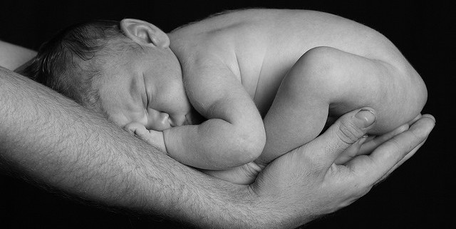
Babies twist, stretch and tumble quite a bit during pregnancy. Before labor starts, however, they usually come to rest in a way that allows them to be delivered through the birth canal headfirst. This position is called cephalic presentation. But there are other ways a baby may settle just before labor begins.
Following are some of the possible ways a baby may be positioned at the end of pregnancy.

Head down, face down
When a baby is head down, face down, the medical term for it is the cephalic occiput anterior position. This the most common position for a baby to be born in. With the face down and turned slightly to the side, the smallest part of the baby's head leads the way through the birth canal. It is the easiest way for a baby to be born.

Head down, face up
When a baby is head down, face up, the medical term for it is the cephalic occiput posterior position. In this position, it might be harder for a baby's head to go under the pubic bone during delivery. That can make labor take longer.
Most babies who begin labor in this position eventually turn to be face down. If that doesn't happen, and the second stage of labor is taking a long time, a member of the health care team may reach through the vagina to help the baby turn. This is called manual rotation.
In some cases, a baby can be born in the head-down, face-up position. Use of forceps or a vacuum device to help with delivery is more common when a baby is in this position than in the head-down, face-down position. In some cases, a C-section delivery may be needed.

Frank breech
When a baby's feet or buttocks are in place to come out first during birth, it's called a breech presentation. This happens in about 3% to 4% of babies close to the time of birth. The baby shown below is in a frank breech presentation. That's when the knees aren't bent, and the feet are close to the baby's head. This is the most common type of breech presentation.
If you are more than 36 weeks into your pregnancy and your baby is in a frank breech presentation, your health care professional may try to move the baby into a head-down position. This is done using a procedure called external cephalic version. It involves one or two members of the health care team putting pressure on your belly with their hands to get the baby to roll into a head-down position.
If the procedure isn't successful, or if the baby moves back into a breech position, talk with a member of your health care team about the choices you have for delivery. Most babies in a frank breech position are born by planned C-section.

Complete and incomplete breech
A complete breech presentation, as shown below, is when the baby has both knees bent and both legs pulled close to the body. In an incomplete breech, one or both of the legs are not pulled close to the body, and one or both of the feet or knees are below the baby's buttocks. If a baby is in either of these positions, you might feel kicking in the lower part of your belly.
If you are more than 36 weeks into your pregnancy and your baby is in a complete or incomplete breech presentation, your health care professional may try to move the baby into a head-down position. This is done using a procedure called external cephalic version. It involves one or two members of the health care team putting pressure on your belly with their hands to get the baby to roll into a head-down position.
If the procedure isn't successful, or if the baby moves back into a breech position, talk with a member of your health care team about the choices you have for delivery. Many babies in a complete or incomplete breech position are born by planned C-section.

When a baby is sideways — lying horizontal across the uterus, rather than vertical — it's called a transverse lie. In this position, the baby's back might be:
- Down, with the back facing the birth canal.
- Sideways, with one shoulder pointing toward the birth canal.
- Up, with the hands and feet facing the birth canal.
Although many babies are sideways early in pregnancy, few stay this way when labor begins.
If your baby is in a transverse lie during week 37 of your pregnancy, your health care professional may try to move the baby into a head-down position. This is done using a procedure called external cephalic version. External cephalic version involves one or two members of your health care team putting pressure on your belly with their hands to get the baby to roll into a head-down position.
If the procedure isn't successful, or if the baby moves back into a transverse lie, talk with a member of your health care team about the choices you have for delivery. Many babies who are in a transverse lie are born by C-section.

If you're pregnant with twins and only the twin that's lower in the uterus is head down, as shown below, your health care provider may first deliver that baby vaginally.
Then, in some cases, your health care team may suggest delivering the second twin in the breech position. Or they may try to move the second twin into a head-down position. This is done using a procedure called external cephalic version. External cephalic version involves one or two members of the health care team putting pressure on your belly with their hands to get the baby to roll into a head-down position.
Your health care team may suggest delivery by C-section for the second twin if:
- An attempt to deliver the baby in the breech position is not successful.
- You do not want to try to have the baby delivered vaginally in the breech position.
- An attempt to move the baby into a head-down position is not successful.
- You do not want to try to move the baby to a head-down position.
In some cases, your health care team may advise that you have both twins delivered by C-section. That might happen if the lower twin is not head down, the second twin has low or high birth weight as compared to the first twin, or if preterm labor starts.

- Landon MB, et al., eds. Normal labor and delivery. In: Gabbe's Obstetrics: Normal and Problem Pregnancies. 8th ed. Elsevier; 2021. https://www.clinicalkey.com. Accessed May 19, 2023.
- Holcroft Argani C, et al. Occiput posterior position. https://www.updtodate.com/contents/search. Accessed May 19, 2023.
- Frequently asked questions: If your baby is breech. American College of Obstetricians and Gynecologists https://www.acog.org/womens-health/faqs/if-your-baby-is-breech. Accessed May 22, 2023.
- Hofmeyr GJ. Overview of breech presentation. https://www.updtodate.com/contents/search. Accessed May 22, 2023.
- Strauss RA, et al. Transverse fetal lie. https://www.updtodate.com/contents/search. Accessed May 22, 2023.
- Chasen ST, et al. Twin pregnancy: Labor and delivery. https://www.updtodate.com/contents/search. Accessed May 22, 2023.
- Cohen R, et al. Is vaginal delivery of a breech second twin safe? A comparison between delivery of vertex and non-vertex second twins. The Journal of Maternal-Fetal & Neonatal Medicine. 2021; doi:10.1080/14767058.2021.2005569.
- Marnach ML (expert opinion). Mayo Clinic. May 31, 2023.
Products and Services
- A Book: Obstetricks
- A Book: Mayo Clinic Guide to a Healthy Pregnancy
- 3rd trimester pregnancy
- Fetal development: The 3rd trimester
- Overdue pregnancy
- Pregnancy due date calculator
- Prenatal care: 3rd trimester
Mayo Clinic does not endorse companies or products. Advertising revenue supports our not-for-profit mission.
- Opportunities
Mayo Clinic Press
Check out these best-sellers and special offers on books and newsletters from Mayo Clinic Press .
- Mayo Clinic on Incontinence - Mayo Clinic Press Mayo Clinic on Incontinence
- The Essential Diabetes Book - Mayo Clinic Press The Essential Diabetes Book
- Mayo Clinic on Hearing and Balance - Mayo Clinic Press Mayo Clinic on Hearing and Balance
- FREE Mayo Clinic Diet Assessment - Mayo Clinic Press FREE Mayo Clinic Diet Assessment
- Mayo Clinic Health Letter - FREE book - Mayo Clinic Press Mayo Clinic Health Letter - FREE book
- Healthy Lifestyle
Make twice the impact
Your gift can go twice as far to advance cancer research and care!
Fetal Presentation, Position, and Lie (Including Breech Presentation)
- Key Points |
Abnormal fetal lie or presentation may occur due to fetal size, fetal anomalies, uterine structural abnormalities, multiple gestation, or other factors. Diagnosis is by examination or ultrasonography. Management is with physical maneuvers to reposition the fetus, operative vaginal delivery , or cesarean delivery .
Terms that describe the fetus in relation to the uterus, cervix, and maternal pelvis are
Fetal presentation: Fetal part that overlies the maternal pelvic inlet; vertex (cephalic), face, brow, breech, shoulder, funic (umbilical cord), or compound (more than one part, eg, shoulder and hand)
Fetal position: Relation of the presenting part to an anatomic axis; for transverse presentation, occiput anterior, occiput posterior, occiput transverse
Fetal lie: Relation of the fetus to the long axis of the uterus; longitudinal, oblique, or transverse
Normal fetal lie is longitudinal, normal presentation is vertex, and occiput anterior is the most common position.
Abnormal fetal lie, presentation, or position may occur with
Fetopelvic disproportion (fetus too large for the pelvic inlet)
Fetal congenital anomalies
Uterine structural abnormalities (eg, fibroids, synechiae)
Multiple gestation
Several common types of abnormal lie or presentation are discussed here.

Transverse lie
Fetal position is transverse, with the fetal long axis oblique or perpendicular rather than parallel to the maternal long axis. Transverse lie is often accompanied by shoulder presentation, which requires cesarean delivery.
Breech presentation
There are several types of breech presentation.
Frank breech: The fetal hips are flexed, and the knees extended (pike position).
Complete breech: The fetus seems to be sitting with hips and knees flexed.
Single or double footling presentation: One or both legs are completely extended and present before the buttocks.
Types of breech presentations
Breech presentation makes delivery difficult ,primarily because the presenting part is a poor dilating wedge. Having a poor dilating wedge can lead to incomplete cervical dilation, because the presenting part is narrower than the head that follows. The head, which is the part with the largest diameter, can then be trapped during delivery.
Additionally, the trapped fetal head can compress the umbilical cord if the fetal umbilicus is visible at the introitus, particularly in primiparas whose pelvic tissues have not been dilated by previous deliveries. Umbilical cord compression may cause fetal hypoxemia.

Predisposing factors for breech presentation include
Preterm labor
Uterine abnormalities
Fetal anomalies
If delivery is vaginal, breech presentation may increase risk of
Umbilical cord prolapse
Birth trauma
Perinatal death

Face or brow presentation
In face presentation, the head is hyperextended, and position is designated by the position of the chin (mentum). When the chin is posterior, the head is less likely to rotate and less likely to deliver vaginally, necessitating cesarean delivery.
Brow presentation usually converts spontaneously to vertex or face presentation.
Occiput posterior position
The most common abnormal position is occiput posterior.
The fetal neck is usually somewhat deflexed; thus, a larger diameter of the head must pass through the pelvis.
Progress may arrest in the second phase of labor. Operative vaginal delivery or cesarean delivery is often required.
Position and Presentation of the Fetus
If a fetus is in the occiput posterior position, operative vaginal delivery or cesarean delivery is often required.
In breech presentation, the presenting part is a poor dilating wedge, which can cause the head to be trapped during delivery, often compressing the umbilical cord.
For breech presentation, usually do cesarean delivery at 39 weeks or during labor, but external cephalic version is sometimes successful before labor, usually at 37 or 38 weeks.

- Cookie Preferences

Copyright © 2024 Merck & Co., Inc., Rahway, NJ, USA and its affiliates. All rights reserved.
Fetal Presentation, Position, and Lie (Including Breech Presentation)
- Key Points |
Abnormal fetal lie or presentation may occur due to fetal size, fetal anomalies, uterine structural abnormalities, multiple gestation, or other factors. Diagnosis is by examination or ultrasonography. Management is with physical maneuvers to reposition the fetus, operative vaginal delivery , or cesarean delivery .
Terms that describe the fetus in relation to the uterus, cervix, and maternal pelvis are
Fetal presentation: Fetal part that overlies the maternal pelvic inlet; vertex (cephalic), face, brow, breech, shoulder, funic (umbilical cord), or compound (more than one part, eg, shoulder and hand)
Fetal position: Relation of the presenting part to an anatomic axis; for transverse presentation, occiput anterior, occiput posterior, occiput transverse
Fetal lie: Relation of the fetus to the long axis of the uterus; longitudinal, oblique, or transverse
Normal fetal lie is longitudinal, normal presentation is vertex, and occiput anterior is the most common position.
Abnormal fetal lie, presentation, or position may occur with
Fetopelvic disproportion (fetus too large for the pelvic inlet)
Fetal congenital anomalies
Uterine structural abnormalities (eg, fibroids, synechiae)
Multiple gestation
Several common types of abnormal lie or presentation are discussed here.
Transverse lie
Fetal position is transverse, with the fetal long axis oblique or perpendicular rather than parallel to the maternal long axis. Transverse lie is often accompanied by shoulder presentation, which requires cesarean delivery.
Breech presentation
There are several types of breech presentation.
Frank breech: The fetal hips are flexed, and the knees extended (pike position).
Complete breech: The fetus seems to be sitting with hips and knees flexed.
Single or double footling presentation: One or both legs are completely extended and present before the buttocks.
Types of breech presentations
Breech presentation makes delivery difficult ,primarily because the presenting part is a poor dilating wedge. Having a poor dilating wedge can lead to incomplete cervical dilation, because the presenting part is narrower than the head that follows. The head, which is the part with the largest diameter, can then be trapped during delivery.
Additionally, the trapped fetal head can compress the umbilical cord if the fetal umbilicus is visible at the introitus, particularly in primiparas whose pelvic tissues have not been dilated by previous deliveries. Umbilical cord compression may cause fetal hypoxemia.
Predisposing factors for breech presentation include
Preterm labor
Uterine abnormalities
Fetal anomalies
If delivery is vaginal, breech presentation may increase risk of
Umbilical cord prolapse
Birth trauma
Perinatal death
Face or brow presentation
In face presentation, the head is hyperextended, and position is designated by the position of the chin (mentum). When the chin is posterior, the head is less likely to rotate and less likely to deliver vaginally, necessitating cesarean delivery.
Brow presentation usually converts spontaneously to vertex or face presentation.
Occiput posterior position
The most common abnormal position is occiput posterior.
The fetal neck is usually somewhat deflexed; thus, a larger diameter of the head must pass through the pelvis.
Progress may arrest in the second phase of labor. Operative vaginal delivery or cesarean delivery is often required.
Position and Presentation of the Fetus
If a fetus is in the occiput posterior position, operative vaginal delivery or cesarean delivery is often required.
In breech presentation, the presenting part is a poor dilating wedge, which can cause the head to be trapped during delivery, often compressing the umbilical cord.
For breech presentation, usually do cesarean delivery at 39 weeks or during labor, but external cephalic version is sometimes successful before labor, usually at 37 or 38 weeks.
- Cookie Preferences
Copyright © 2024 Merck & Co., Inc., Rahway, NJ, USA and its affiliates. All rights reserved.

An official website of the United States government
The .gov means it's official. Federal government websites often end in .gov or .mil. Before sharing sensitive information, make sure you're on a federal government site.
The site is secure. The https:// ensures that you are connecting to the official website and that any information you provide is encrypted and transmitted securely.
- Publications
- Account settings
- Browse Titles
NCBI Bookshelf. A service of the National Library of Medicine, National Institutes of Health.
StatPearls [Internet]. Treasure Island (FL): StatPearls Publishing; 2024 Jan-.

StatPearls [Internet].
Delivery, face and brow presentation.
Julija Makajeva ; Mohsina Ashraf .
Affiliations
Last Update: January 9, 2023 .
- Continuing Education Activity
Face and brow presentation is a malpresentation during labor when the presenting part is either the face or, in the case of brow presentation, it is the area between the orbital ridge and the anterior fontanelle. This activity reviews the evaluation and management of these two presentations and explains the role of the interprofessional team in managing delivery safely for both the mother and the baby.
- Describe the mechanism of labor in the face and brow presentation.
- Summarize potential maternal and fetal complications during the face and brow presentations.
- Review different management approaches for the face and brow presentation.
- Outline some interprofessional strategies that will improve patient outcomes in delivery cases with face and brow presentation issues.
- Introduction
The term presentation describes the leading part of the fetus or the anatomical structure closest to the maternal pelvic inlet during labor. The presentation can roughly be divided into the following classifications: cephalic, breech, shoulder, and compound. Cephalic presentation is the most common and can be further subclassified as vertex, sinciput, brow, face, and chin. The most common presentation in term labor is the vertex, where the fetal neck is flexed to the chin, minimizing the head circumference.
Face presentation – an abnormal form of cephalic presentation where the presenting part is mentum. This typically occurs because of hyperextension of the neck and the occiput touching the fetal back. Incidence of face presentation is rare, accounting for approximately 1 in 600 of all presentations. [1] [2] [3]
In brow presentation, the neck is not extended as much as in face presentation, and the leading part is the area between the anterior fontanelle and the orbital ridges. Brow presentation is considered the rarest of all malpresentation with a prevalence of 1 in 500 to 1 in 4000 deliveries. [3]
Both face and brow presentations occur due to extension of the fetal neck instead of flexion; therefore, conditions that would lead to hyperextension or prevent flexion of the fetal neck can all contribute to face or brow presentation. These risk factors may be related to either the mother or the fetus. Maternal risk factors are preterm delivery, contracted maternal pelvis, platypelloid pelvis, multiparity, previous cesarean section, black race. Fetal risk factors include anencephaly, multiple loops of cord around the neck, masses of the neck, macrosomia, polyhydramnios. [2] [4] [5]
These malpresentations are usually diagnosed during the second stage of labor when performing a digital examination. It is possible to palpate orbital ridges, nose, malar eminences, mentum, mouth, gums, and chin in face presentation. Based on the position of the chin, face presentation can be further divided into mentum anterior, posterior, or transverse. In brow presentation, anterior fontanelle and face can be palpated except for the mouth and the chin. Brow presentation can then be further described based on the position of the anterior fontanelle as frontal anterior, posterior, or transverse.
Diagnosing the exact presentation can be challenging, and face presentation may be misdiagnosed as frank breech. To avoid any confusion, a bedside ultrasound scan can be performed. [6] The ultrasound imaging can show a reduced angle between the occiput and the spine or, the chin is separated from the chest. However, ultrasound does not provide much predicting value in the outcome of the labor. [7]
- Anatomy and Physiology
Before discussing the mechanism of labor in the face or brow presentation, it is crucial to highlight some anatomical landmarks and their measurements.
Planes and Diameters of the Pelvis
The three most important planes in the female pelvis are the pelvic inlet, mid pelvis, and pelvic outlet.
Four diameters can describe the pelvic inlet: anteroposterior, transverse, and two obliques. Furthermore, based on the different landmarks on the pelvic inlet, there are three different anteroposterior diameters, named conjugates: true conjugate, obstetrical conjugate, and diagonal conjugate. Only the latter can be measured directly during the obstetric examination. The shortest of these three diameters is obstetrical conjugate, which measures approximately 10.5 cm and is a distance between the sacral promontory and 1 cm below the upper border of the symphysis pubis. This measurement is clinically significant as the fetal head must pass through this diameter during the engagement phase. The transverse diameter measures about 13.5cm and is the widest distance between the innominate line on both sides.
The shortest distance in the mid pelvis is the interspinous diameter and usually is only about 10 cm.
Fetal Skull Diameters
There are six distinguished longitudinal fetal skull diameters:
- Suboccipito-bregmatic: from the center of anterior fontanelle (bregma) to the occipital protuberance, measuring 9.5 cm. This is the presenting diameter in vertex presentation.
- Suboccipito-frontal: from the anterior part of bregma to the occipital protuberance, measuring 10 cm
- Occipito-frontal: from the root of the nose to the most prominent part of the occiput, measuring 11.5cm
- Submento-bregmatic: from the center of the bregma to the angle of the mandible, measuring 9.5 cm. This is the presenting diameter in face presentation where the neck is hyperextended.
- Submento-vertical: from the midpoint between fontanelles and the angle of the mandible, measuring 11.5cm
- Occipito-mental: from the midpoint between fontanelles and the tip of the chin, measuring 13.5 cm. It is the presenting diameter in brow presentation.
Cardinal Movements of Normal Labor
- Neck flexion
- Internal rotation
- Extension (delivers head)
- External rotation (Restitution)
- Expulsion (delivery of anterior and posterior shoulders)
Some of the key movements are not possible in the face or brow presentations.
Based on the information provided above, it is obvious that labor will be arrested in brow presentation unless it spontaneously changes to face or vertex, as the occipito-mental diameter of the fetal head is significantly wider than the smallest diameter of the female pelvis. Face presentation can, however, be delivered vaginally, and further mechanisms of face delivery will be explained in later sections.
- Indications
As mentioned previously, spontaneous vaginal delivery can be successful in face presentation. However, the main indication for vaginal delivery in such circumstances would be a maternal choice. It is crucial to have a thorough conversation with a mother, explaining the risks and benefits of vaginal delivery with face presentation and a cesarean section. Informed consent and creating a rapport with the mother is an essential aspect of safe and successful labor.
- Contraindications
Vaginal delivery of face presentation is contraindicated if the mentum is lying posteriorly or is in a transverse position. In such a scenario, the fetal brow is pressing against the maternal symphysis pubis, and the short fetal neck, which is already maximally extended, cannot span the surface of the maternal sacrum. In this position, the diameter of the head is larger than the maternal pelvis, and it cannot descend through the birth canal. Therefore the cesarean section is recommended as the safest mode of delivery for mentum posterior face presentations.
Attempts to manually convert face presentation to vertex, manual or forceps rotation of the persistent posterior chin to anterior are contraindicated as they can be dangerous.
Persistent brow presentation itself is a contraindication for vaginal delivery unless the fetus is significantly small or the maternal pelvis is large.
Continuous electronic fetal heart rate monitoring is recommended for face and brow presentations, as heart rate abnormalities are common in these scenarios. One study found that only 14% of the cases with face presentation had no abnormal traces on the cardiotocograph. [8] It is advised to use external transducer devices to prevent damage to the eyes. When internal monitoring is inevitable, it is suggested to place monitoring devices on bony parts carefully.
People who are usually involved in the delivery of face/ brow presentation are:
- Experienced midwife, preferably looking after laboring woman 1:1
- Senior obstetrician
- Neonatal team - in case of need for resuscitation
- Anesthetic team - to provide necessary pain control (e.g., epidural)
- Theatre team - in case of failure to progress and an emergency cesarean section will be required.
- Preparation
No specific preparation is required for face or brow presentation. However, it is essential to discuss the labor options with the mother and birthing partner and inform members of the neonatal, anesthetic, and theatre co-ordinating teams.
- Technique or Treatment
Mechanism of Labor in Face Presentation
During contractions, the pressure exerted by the fundus of the uterus on the fetus and pressure of amniotic fluid initiate descent. During this descent, the fetal neck extends instead of flexing. The internal rotation determines the outcome of delivery, if the fetal chin rotates posteriorly, vaginal delivery would not be possible, and cesarean section is permitted. The approach towards mentum-posterior delivery should be individualized, as the cases are rare. Expectant management is acceptable in multiparous women with small fetuses, as a spontaneous mentum-anterior rotation can occur. However, there should be a low threshold for cesarean section in primigravida women or women with large fetuses.
When the fetal chin is rotated towards maternal symphysis pubis as described as mentum-anterior; in these cases further descend through the vaginal canal continues with approximately 73% cases deliver spontaneously. [9] Fetal mentum presses on the maternal symphysis pubis, and the head is delivered by flexion. The occiput is pointing towards the maternal back, and external rotation happens. Shoulders are delivered in the same manner as in vertex delivery.
Mechanism of Labor in Brow Presentation
As this presentation is considered unstable, it is usually converted into a face or an occiput presentation. Due to the cephalic diameter being wider than the maternal pelvis, the fetal head cannot engage; thus, brow delivery cannot take place. Unless the fetus is small or the pelvis is very wide, the prognosis for vaginal delivery is poor. With persistent brow presentation, a cesarean section is required for safe delivery.
- Complications
As the cesarean section is becoming a more accessible mode of delivery in malpresentations, the incidence of maternal and fetal morbidity and mortality during face presentation has dropped significantly. [10]
However, there are still some complications associated with the nature of labor in face presentation. Due to the fetal head position, it is more challenging for the head to engage in the birth canal and descend, resulting in prolonged labor.
Prolonged labor itself can provoke foetal distress and arrhythmias. If the labor arrests or signs of fetal distress appear on CTG, the recommended next step in management is an emergency cesarean section, which in itself carries a myriad of operative and post-operative complications.
Finally, due to the nature of the fetal position and prolonged duration of labor in face presentation, neonates develop significant edema of the skull and face. Swelling of the fetal airway may also be present, resulting in respiratory distress after birth and possible intubation.
- Clinical Significance
During vertex presentation, the fetal head flexes, bringing the chin to the chest, forming the smallest possible fetal head diameter, measuring approximately 9.5cm. With face and brow presentation, the neck hyperextends, resulting in greater cephalic diameters. As a result, the fetal head will engage later, and labor will progress more slowly. Failure to progress in labor is also more common in both presentations compared to vertex presentation.
Furthermore, when the fetal chin is in a posterior position, this prevents further flexion of the fetal neck, as browns are pressing on the symphysis pubis. As a result, descend through the birth canal is impossible. Such presentation is considered undeliverable vaginally and requires an emergency cesarean section.
Manual attempts to change face presentation to vertex, manual or forceps rotation to mentum anterior are considered dangerous and are discouraged.
- Enhancing Healthcare Team Outcomes
A multidisciplinary team of healthcare experts supports the woman and her child during labor and the perinatal period. For a face or brow presentation to be appropriately diagnosed, an experienced midwife and obstetrician must be involved in the vaginal examination and labor monitoring. As fetal anomalies, such as anencephaly or goiter, can contribute to face presentation, sonographers experienced in antenatal scanning should also be involved in the care. It is advised to inform the anesthetic and neonatal teams in advance of the possible need for emergency cesarean section and resuscitation of the neonate. [11] [12]
- Review Questions
- Access free multiple choice questions on this topic.
- Comment on this article.
Disclosure: Julija Makajeva declares no relevant financial relationships with ineligible companies.
Disclosure: Mohsina Ashraf declares no relevant financial relationships with ineligible companies.
This book is distributed under the terms of the Creative Commons Attribution-NonCommercial-NoDerivatives 4.0 International (CC BY-NC-ND 4.0) ( http://creativecommons.org/licenses/by-nc-nd/4.0/ ), which permits others to distribute the work, provided that the article is not altered or used commercially. You are not required to obtain permission to distribute this article, provided that you credit the author and journal.
- Cite this Page Makajeva J, Ashraf M. Delivery, Face and Brow Presentation. [Updated 2023 Jan 9]. In: StatPearls [Internet]. Treasure Island (FL): StatPearls Publishing; 2024 Jan-.
In this Page
Bulk download.
- Bulk download StatPearls data from FTP
Related information
- PubMed Links to PubMed
Similar articles in PubMed
- Sonographic diagnosis of fetal head deflexion and the risk of cesarean delivery. [Am J Obstet Gynecol MFM. 2020] Sonographic diagnosis of fetal head deflexion and the risk of cesarean delivery. Bellussi F, Livi A, Cataneo I, Salsi G, Lenzi J, Pilu G. Am J Obstet Gynecol MFM. 2020 Nov; 2(4):100217. Epub 2020 Aug 18.
- Review Sonographic evaluation of the fetal head position and attitude during labor. [Am J Obstet Gynecol. 2022] Review Sonographic evaluation of the fetal head position and attitude during labor. Ghi T, Dall'Asta A. Am J Obstet Gynecol. 2022 Jul 6; . Epub 2022 Jul 6.
- Stages of Labor. [StatPearls. 2024] Stages of Labor. Hutchison J, Mahdy H, Hutchison J. StatPearls. 2024 Jan
- Leopold Maneuvers. [StatPearls. 2024] Leopold Maneuvers. Superville SS, Siccardi MA. StatPearls. 2024 Jan
- Review Labor with abnormal presentation and position. [Obstet Gynecol Clin North Am. ...] Review Labor with abnormal presentation and position. Stitely ML, Gherman RB. Obstet Gynecol Clin North Am. 2005 Jun; 32(2):165-79.
Recent Activity
- Delivery, Face and Brow Presentation - StatPearls Delivery, Face and Brow Presentation - StatPearls
Your browsing activity is empty.
Activity recording is turned off.
Turn recording back on
Connect with NLM
National Library of Medicine 8600 Rockville Pike Bethesda, MD 20894
Web Policies FOIA HHS Vulnerability Disclosure
Help Accessibility Careers
Fetal Presentation: Baby’s First Pose
Share this post

Occiput Anterior
Occiput posterior, transverse position, complete breech, frank breech, changing fetal presentation, baby positions.
The position in which your baby develops is called the “fetal presentation.” During most of your pregnancy, the baby will be curled up in a ball – that’s why we call it the “fetal position.” The baby might flip around over the course of development, which is why you can sometimes feel a foot poking into your side or an elbow prodding your bellybutton. As you get closer to delivery, the baby will change positions and move lower in your uterus in preparation. Over the last part of your pregnancy, your doctor or medical care provider will monitor the baby’s position to keep an eye out for any potential problems.
In the occiput anterior position, the baby is pointed headfirst toward the birth canal and is facing down – toward your back. This is the easiest possible position for delivery because it allows the crown of the baby’s head to pass through first, followed by the shoulders and the rest of the body. The crown of the head is the narrowest part, so it can lead the way for the rest of the head.
The baby’s head will move slowly downward as you get closer to delivery until it “engages” with your pelvis. At that point, the baby’s head will fit snugly and won’t be able to wobble around. That’s exactly where you want to be just before labor. The occiput anterior position causes the least stress on your little one and the easiest labor for you.
In the occiput posterior position, the baby is pointed headfirst toward the birth canal but is facing upward, toward your stomach. This can trap the baby’s head under your pubic bone, making it harder to get out through the birth canal. In most cases, a baby in the occiput posterior position will either turn around naturally during the course of labor or your doctor or midwife may help it along manually or with forceps.
In a transverse position, the baby is sideways across the birth canal rather than head- or feet-first. It’s rare for a baby to stay in this position all the way up to delivery, but your doctor may attempt to gently push on your abdomen until the baby is in a more favorable fetal presentation. If you go into labor while the baby is in a transverse position, your medical care provider will likely recommend a c-section to avoid stressing or injuring the baby.
Breech Presentation
If the baby’s legs or buttocks are leading the way instead of the head, it’s called a breech presentation. It’s much harder to deliver in this position – the baby’s limbs are unlikely to line up all in the right direction and the birth canal likely won’t be stretched enough to allow the head to pass. Breech presentation used to be extremely dangerous for mothers and children both, and it’s still not easy, but medical intervention can help.
Sometimes, the baby will turn around and you’ll be able to deliver vaginally. Most healthcare providers, however, recommend a cesarean section for all breech babies because of the risks of serious injury to both mother and child in a breech vaginal delivery.
A complete breech position refers to the baby being upside down for delivery – feet first and head up. The baby’s legs are folded up and the feet are near the buttocks.
In a frank breech position, the baby’s legs are extended and the baby’s buttocks are closest to the birth canal. This is the most common breech presentation .
By late in your pregnancy, your baby can already move around – you’re probably feeling those kicks! Unfortunately, your little one doesn’t necessarily know how to aim for the birth canal. If the baby isn’t in the occiput anterior position by about 32 weeks, your doctor or midwife will typically recommend trying adjust the fetal presentation. They’ll use monitors to keep an eye on the baby and watch for signs of stress as they push and lift on your belly to coax your little one into the right spot. Your doctor may also advise you to try certain exercises at home to encourage the baby to move into the proper position. For example, getting on your hands and knees for a few minutes every day can help bring the baby around. You can also put cushions on your chairs to make sure your hips are always elevated, which can help move things into the right place. It’s important to start working on the proper fetal position early, as it becomes much harder to adjust after about 37 weeks when there’s less room to move around.
In many cases, the baby will eventually line up properly before delivery. Sometimes, however, the baby is still in the wrong spot by the time you go into labor. Your doctor or midwife may be able to move the baby during labor using forceps or ventouse . If that’s not possible, it’s generally safer for you and the baby if you deliver by c-section.
Image Credit and License
Leave a Reply
Your email address will not be published. Required fields are marked *
Save my name, email, and website in this browser for the next time I comment.
- Stages of Pregnancy
- Foods to Avoid
- Medicines to Avoid
- Pregnancy Road Map
Birth Injuries
- Cerebral Palsy
- Brachial Plexus Injuries & Erb’s Palsy
- Brain Damage
- Meconium Aspiration
- Bone Fractures
- Nerve Damage
Newborn Care
- Baby Development
Legal Issues
- Birth Injury vs. Birth Defect
- Birth Injury Lawsuits
- Proving Your Case
- Elements Of A Case

- Email Address *
- Phone Number *
- Comments This field is for validation purposes and should be left unchanged.
- Trying to Conceive
- Signs & Symptoms
- Pregnancy Tests
- Fertility Testing
- Fertility Treatment
- Weeks & Trimesters
- Staying Healthy
- Preparing for Baby
- Complications & Concerns
- Pregnancy Loss
- Breastfeeding
- School-Aged Kids
- Raising Kids
- Personal Stories
- Everyday Wellness
- Safety & First Aid
- Immunizations
- Food & Nutrition
- Active Play
- Pregnancy Products
- Nursery & Sleep Products
- Nursing & Feeding Products
- Clothing & Accessories
- Toys & Gifts
- Ovulation Calculator
- Pregnancy Due Date Calculator
- How to Talk About Postpartum Depression
- Editorial Process
- Meet Our Review Board
Fetal Positions for Labor and Birth
Knowing your baby's position can you help ease pain and speed up labor
In the last weeks of pregnancy , determining your baby's position can help you manage pain and discomfort. Knowing your baby's position during early labor can help you adjust your own position during labor and possibly even speed up the process.
Right or Left Occiput Anterior
Illustration by JR Bee, Verywell
Looking at where the baby's head is in the birth canal helps determine the fetal position.The front of a baby's head is referred to as the anterior portion and the back is the posterior portion. There are two different positions called occiput anterior (OA) positions that may occur.
The left occiput anterior (LOA) position is the most common in labor. In this position, the baby's head is slightly off-center in the pelvis with the back of the head toward the mother's left thigh.
The right occiput anterior (ROA) presentation is also common in labor. In this position, the back of the baby is slightly off-center in the pelvis with the back of the head toward the mother's right thigh.
In general, OA positions do not lead to problems or additional pain during labor or birth.
Right or Left Occiput Transverse
Illustration by JR Bee, Verywell
When facing out toward the mother's right thigh, the baby is said to be left occiput transverse (LOT). This position is halfway between a posterior and anterior position. If the baby was previously in a posterior position (in either direction), the LOT position indicates positive movement toward an anterior position.
When the baby is facing outward toward the mother's left thigh, the baby is said to be right occiput transverse (ROT). Like the previous presentation, ROT is halfway between a posterior and anterior position. If the baby was previously in a posterior position, ROT is a sign the baby is making a positive move toward an anterior position.
When a baby is in the left occiput transverse position (LOT) or right occiput transverse (ROT) position during labor, it may lead to more pain and a slower progression.
Tips to Reduce Discomfort
There are several labor positions a mother can try to alleviate pain and encourage the baby to continue rotating toward an anterior position, including:
- Pelvic tilts
- Standing and swaying
A doula , labor nurse, midwife , or doctor may have other suggestions for positions.
Right or Left Occiput Posterior
When facing forward, the baby is in the occiput posterior position. If the baby is facing forward and slightly to the left (looking toward the mother's right thigh) it is in the left occiput posterior (LOP) position. This presentation can lead to more back pain (sometimes referred to as " back labor ") and slow progression of labor.
In the right occiput posterior position (ROP), the baby is facing forward and slightly to the right (looking toward the mother's left thigh). This presentation may slow labor and cause more pain.
To help prevent or decrease pain during labor and encourage the baby to move into a better position for delivery, mothers can try a variety of positions, including:
- Hands and knees
- Pelvic rocking
Mothers may try other comfort measures, including:
- Bathtub or shower (water)
- Counter pressure
- Movement (swaying, dancing, sitting on a birth ball )
- Rice socks (heat packs)
How a Doctor Determines Baby's Position
Leopold's maneuvers are a series of hands-on examinations your doctor or midwife will use to help determine your baby's position. During the third trimester , the assessment will be done at most of your prenatal visits. Knowing the baby's position before labor begins can help you prepare for labor and delivery.
Once labor begins, a nurse, doctor, or midwife will be able to get a more accurate sense of your baby's position by performing a vaginal exam. When your cervix is dilated enough, the practitioner will insert their fingers into the vagina and feel for the suture lines of the baby's skull as it moves down in the birth canal. It's important to ensure the baby is head down and moving in the right direction.
Labor and delivery may be more complicated if the baby is not in a head-down position, such as in the case of a breech presentation.
How You Can Determine Baby's Position
While exams by health practitioners are an important part of your care, from the prenatal period through labor and delivery, often the best person to assess a baby's position in the pelvis is you. Mothers should pay close attention to how the baby moves and where different movements are felt.
A technique called belly mapping can help mothers ask questions of themselves to assess their baby's movement and get a sense of the position they are in as labor approaches.
For example, the position of your baby's legs can be determined by asking questions about the location and strength of the kicking you feel. The spots where you feel the strongest kicks are most likely where your baby's feet are.
Other landmarks you can feel for include a large, flat plane, which is most likely your baby's back. Sometimes you can feel the baby arching his or her back.
At the top or bottom of the flat plane, you may feel either a hard, round shape (most likely your baby's head) or a soft curve (most likely to be your baby's bottom).
Guittier M, Othenin-Girard V, de Gasquet B, Irion O, Boulvain M. Maternal positioning to correct occiput posterior fetal position during the first stage of labour: a randomised controlled trial . BJOG: An International Journal of Obstetrics & Gynaecology . 2016;123(13):2199-2207. doi:10.1111/1471-0528.13855
Gizzo S, Di Gangi S, Noventa M, Bacile V, Zambon A, Nardelli G. Women’s Choice of Positions during Labour: Return to the Past or a Modern Way to Give Birth? A Cohort Study in Italy . Biomed Res Int . 2014;2014:1-7. doi:10.1155/2014/638093
Ahmad A, Webb S, Early B, Sitch A, Khan K, MacArthur C. Association between fetal position at onset of labor and mode of delivery: a prospective cohort study . Ultrasound in Obstetrics & Gynecology . 2014;43(2):176-182. doi:10.1002/uog.13189
Nishikawa M, Sakakibara H. Effect of nursing intervention program using abdominal palpation of Leopold’s maneuvers on maternal-fetal attachment . Reprod Health . 2013;10(1). doi:10.1186/1742-4755-10-12
Choi S, Park Y, Lee D, Ko H, Park I, Shin J. Sonographic assessment of fetal occiput position during labor for the prediction of labor dystocia and perinatal outcomes . The Journal of Maternal-Fetal & Neonatal Medicine . 2016;29(24):3988-3992. doi:10.3109/14767058.2016.1152250
Bamberg C, Deprest J, Sindhwani N et al. Evaluating fetal head dimension changes during labor using open magnetic resonance imaging . J Perinat Med . 2017;45(3). doi:10.1515/jpm-2016-0005
Gabbe S, Niebyl J, Simpson J et al. Obstetrics . Philadelphia, Pa.: Elsevier; 2012.
By Robin Elise Weiss, PhD, MPH Robin Elise Weiss, PhD, MPH is a professor, author, childbirth and postpartum educator, certified doula, and lactation counselor.
Learn how UpToDate can help you.
Select the option that best describes you
- Medical Professional
- Resident, Fellow, or Student
- Hospital or Institution
- Group Practice
- Patient or Caregiver
- Find in topic
RELATED TOPICS
INTRODUCTION
PATHOGENESIS AND RISK FACTORS
● The fetus does not fully occupy the pelvis, thus allowing a fetal extremity room to prolapse. Predisposing factors include early gestational age, multiple gestation, polyhydramnios, or a large maternal pelvis relative to fetal size [ 2,3 ].
● Membrane rupture occurs when the presenting part is still high, which allows flow of amniotic fluid to carry a fetal extremity, umbilical cord, or both toward the birth canal.
- Pregnancy Classes

Breech Births
In the last weeks of pregnancy, a baby usually moves so his or her head is positioned to come out of the vagina first during birth. This is called a vertex presentation. A breech presentation occurs when the baby’s buttocks, feet, or both are positioned to come out first during birth. This happens in 3–4% of full-term births.
What are the different types of breech birth presentations?
- Complete breech: Here, the buttocks are pointing downward with the legs folded at the knees and feet near the buttocks.
- Frank breech: In this position, the baby’s buttocks are aimed at the birth canal with its legs sticking straight up in front of his or her body and the feet near the head.
- Footling breech: In this position, one or both of the baby’s feet point downward and will deliver before the rest of the body.
What causes a breech presentation?
The causes of breech presentations are not fully understood. However, the data show that breech birth is more common when:
- You have been pregnant before
- In pregnancies of multiples
- When there is a history of premature delivery
- When the uterus has too much or too little amniotic fluid
- When there is an abnormally shaped uterus or a uterus with abnormal growths, such as fibroids
- The placenta covers all or part of the opening of the uterus placenta previa
How is a breech presentation diagnosed?
A few weeks prior to the due date, the health care provider will place her hands on the mother’s lower abdomen to locate the baby’s head, back, and buttocks. If it appears that the baby might be in a breech position, they can use ultrasound or pelvic exam to confirm the position. Special x-rays can also be used to determine the baby’s position and the size of the pelvis to determine if a vaginal delivery of a breech baby can be safely attempted.
Can a breech presentation mean something is wrong?
Even though most breech babies are born healthy, there is a slightly elevated risk for certain problems. Birth defects are slightly more common in breech babies and the defect might be the reason that the baby failed to move into the right position prior to delivery.
Can a breech presentation be changed?
It is preferable to try to turn a breech baby between the 32nd and 37th weeks of pregnancy . The methods of turning a baby will vary and the success rate for each method can also vary. It is best to discuss the options with the health care provider to see which method she recommends.
Medical Techniques
External Cephalic Version (EVC) is a non-surgical technique to move the baby in the uterus. In this procedure, a medication is given to help relax the uterus. There might also be the use of an ultrasound to determine the position of the baby, the location of the placenta and the amount of amniotic fluid in the uterus.
Gentle pushing on the lower abdomen can turn the baby into the head-down position. Throughout the external version the baby’s heartbeat will be closely monitored so that if a problem develops, the health care provider will immediately stop the procedure. ECV usually is done near a delivery room so if a problem occurs, a cesarean delivery can be performed quickly. The external version has a high success rate and can be considered if you have had a previous cesarean delivery.
ECV will not be tried if:
- You are carrying more than one fetus
- There are concerns about the health of the fetus
- You have certain abnormalities of the reproductive system
- The placenta is in the wrong place
- The placenta has come away from the wall of the uterus ( placental abruption )
Complications of EVC include:
- Prelabor rupture of membranes
- Changes in the fetus’s heart rate
- Placental abruption
- Preterm labor
Vaginal delivery versus cesarean for breech birth?
Most health care providers do not believe in attempting a vaginal delivery for a breech position. However, some will delay making a final decision until the woman is in labor. The following conditions are considered necessary in order to attempt a vaginal birth:
- The baby is full-term and in the frank breech presentation
- The baby does not show signs of distress while its heart rate is closely monitored.
- The process of labor is smooth and steady with the cervix widening as the baby descends.
- The health care provider estimates that the baby is not too big or the mother’s pelvis too narrow for the baby to pass safely through the birth canal.
- Anesthesia is available and a cesarean delivery possible on short notice
What are the risks and complications of a vaginal delivery?
In a breech birth, the baby’s head is the last part of its body to emerge making it more difficult to ease it through the birth canal. Sometimes forceps are used to guide the baby’s head out of the birth canal. Another potential problem is cord prolapse . In this situation the umbilical cord is squeezed as the baby moves toward the birth canal, thus slowing the baby’s supply of oxygen and blood. In a vaginal breech delivery, electronic fetal monitoring will be used to monitor the baby’s heartbeat throughout the course of labor. Cesarean delivery may be an option if signs develop that the baby may be in distress.
When is a cesarean delivery used with a breech presentation?
Most health care providers recommend a cesarean delivery for all babies in a breech position, especially babies that are premature. Since premature babies are small and more fragile, and because the head of a premature baby is relatively larger in proportion to its body, the baby is unlikely to stretch the cervix as much as a full-term baby. This means that there might be less room for the head to emerge.
Want to Know More?
- Creating Your Birth Plan
- Labor & Birth Terms to Know
- Cesarean Birth After Care
Compiled using information from the following sources:
- ACOG: If Your Baby is Breech
- William’s Obstetrics Twenty-Second Ed. Cunningham, F. Gary, et al, Ch. 24.
- Danforth’s Obstetrics and Gynecology Ninth Ed. Scott, James R., et al, Ch. 21.
BLOG CATEGORIES
- Can I get pregnant if… ? 3
- Child Adoption 19
- Fertility 54
- Pregnancy Loss 11
- Breastfeeding 29
- Changes In Your Body 5
- Cord Blood 4
- Genetic Disorders & Birth Defects 17
- Health & Nutrition 2
- Is it Safe While Pregnant 54
- Labor and Birth 65
- Multiple Births 10
- Planning and Preparing 24
- Pregnancy Complications 68
- Pregnancy Concerns 62
- Pregnancy Health and Wellness 149
- Pregnancy Products & Tests 8
- Pregnancy Supplements & Medications 14
- The First Year 41
- Week by Week Newsletter 40
- Your Developing Baby 16
- Options for Unplanned Pregnancy 18
- Paternity Tests 2
- Pregnancy Symptoms 5
- Prenatal Testing 16
- The Bumpy Truth Blog 7
- Uncategorized 4
- Abstinence 3
- Birth Control Pills, Patches & Devices 21
- Women's Health 34
- Thank You for Your Donation
- Unplanned Pregnancy
- Getting Pregnant
- Healthy Pregnancy
- Privacy Policy
Share this post:
Similar post.

Episiotomy: Advantages & Complications

Retained Placenta

What is Dilation in Pregnancy?
Track your baby’s development, subscribe to our week-by-week pregnancy newsletter.
- The Bumpy Truth Blog
- Fertility Products Resource Guide
Pregnancy Tools
- Ovulation Calendar
- Baby Names Directory
- Pregnancy Due Date Calculator
- Pregnancy Quiz
Pregnancy Journeys
- Partner With Us
- Corporate Sponsors
- Mammary Glands
- Fallopian Tubes
- Supporting Ligaments
- Reproductive System
- Gametogenesis
- Placental Development
- Maternal Adaptations
- Menstrual Cycle
- Antenatal Care
- Small for Gestational Age
- Large for Gestational Age
- RBC Isoimmunisation
- Prematurity
- Prolonged Pregnancy
- Multiple Pregnancy
- Miscarriage
- Recurrent Miscarriage
- Ectopic Pregnancy
- Hyperemesis Gravidarum
- Gestational Trophoblastic Disease
- Breech Presentation
- Abnormal lie, Malpresentation and Malposition
- Oligohydramnios
- Polyhydramnios
- Placenta Praevia
- Placental Abruption
- Pre-Eclampsia
- Gestational Diabetes
- Headaches in Pregnancy
- Haematological
- Obstetric Cholestasis
- Thyroid Disease in Pregnancy
- Epilepsy in Pregnancy
- Induction of Labour
- Operative Vaginal Delivery
- Prelabour Rupture of Membranes
- Caesarean Section
- Shoulder Dystocia
- Cord Prolapse
- Uterine Rupture
- Amniotic Fluid Embolism
- Primary PPH
- Secondary PPH
- Psychiatric Disease
- Postpartum Contraception
- Breastfeeding Problems
- Primary Dysmenorrhoea
- Amenorrhoea and Oligomenorrhoea
- Heavy Menstrual Bleeding
- Endometriosis
- Endometrial Cancer
- Adenomyosis
- Cervical Polyps
- Cervical Ectropion
- Cervical Intraepithelial Neoplasia + Cervical Screening
- Cervical Cancer
- Polycystic Ovary Syndrome (PCOS)
- Ovarian Cysts & Tumours
- Urinary Incontinence
- Genitourinary Prolapses
- Bartholin's Cyst
- Lichen Sclerosus
- Vulval Carcinoma
- Introduction to Infertility
- Female Factor Infertility
- Male Factor Infertility
- Female Genital Mutilation
- Barrier Contraception
- Combined Hormonal
- Progesterone Only Hormonal
- Intrauterine System & Device
- Emergency Contraception
- Pelvic Inflammatory Disease
- Genital Warts
- Genital Herpes
- Trichomonas Vaginalis
- Bacterial Vaginosis
- Vulvovaginal Candidiasis
- Obstetric History
- Gynaecological History
- Sexual History
- Obstetric Examination
- Speculum Examination
- Bimanual Examination
- Amniocentesis
- Chorionic Villus Sampling
- Hysterectomy
- Endometrial Ablation
- Tension-Free Vaginal Tape
- Contraceptive Implant
- Fitting an IUS or IUD
Abnormal Fetal lie, Malpresentation and Malposition
Original Author(s): Anna Mcclune Last updated: 1st December 2018 Revisions: 12
- 1 Definitions
- 2 Risk Factors
- 3.2 Presentation
- 3.3 Position
- 4 Investigations
- 5.1 Abnormal Fetal Lie
- 5.2 Malpresentation
- 5.3 Malposition
The lie, presentation and position of a fetus are important during labour and delivery.
In this article, we will look at the risk factors, examination and management of abnormal fetal lie, malpresentation and malposition.
Definitions
- Longitudinal, transverse or oblique
- Cephalic vertex presentation is the most common and is considered the safest
- Other presentations include breech, shoulder, face and brow
- Usually the fetal head engages in the occipito-anterior position (the fetal occiput facing anteriorly) – this is ideal for birth
- Other positions include occipito-posterior and occipito-transverse.
Note: Breech presentation is the most common malpresentation, and is covered in detail here .

Fig 1 – The two most common fetal presentations: cephalic and breech.
Risk Factors
The risk factors for abnormal fetal lie, malpresentation and malposition include:
- Multiple pregnancy
- Uterine abnormalities (e.g fibroids, partial septate uterus)
- Fetal abnormalities
- Placenta praevia
- Primiparity
Identifying Fetal Lie, Presentation and Position
The fetal lie and presentation can usually be identified via abdominal examination. The fetal position is ascertained by vaginal examination.
For more information on the obstetric examination, see here .
- Face the patient’s head
- Place your hands on either side of the uterus and gently apply pressure; one side will feel fuller and firmer – this is the back, and fetal limbs may feel ‘knobbly’ on the opposite side
Presentation
- Palpate the lower uterus (above the symphysis pubis) with the fingers of both hands; the head feels hard and round (cephalic) and the bottom feels soft and triangular (breech)
- You may be able to gently push the fetal head from side to side
The fetal lie and presentation may not be possible to identify if the mother has a high BMI, if she has not emptied her bladder, if the fetus is small or if there is polyhydramnios .
During labour, vaginal examination is used to assess the position of the fetal head (in a cephalic vertex presentation). The landmarks of the fetal head, including the anterior and posterior fontanelles, indicate the position.

Fig 2 – Assessing fetal lie and presentation.
Investigations
Any suspected abnormal fetal lie or malpresentation should be confirmed by an ultrasound scan . This could also demonstrate predisposing uterine or fetal abnormalities.
Abnormal Fetal Lie
If the fetal lie is abnormal, an external cephalic version (ECV) can be attempted – ideally between 36 and 38 weeks gestation.
ECV is the manipulation of the fetus to a cephalic presentation through the maternal abdomen.
It has an approximate success rate of 50% in primiparous women and 60% in multiparous women. Only 8% of breech presentations will spontaneously revert to cephalic in primiparous women over 36 weeks gestation.
Complications of ECV are rare but include fetal distress , premature rupture of membranes, antepartum haemorrhage (APH) and placental abruption. The risk of an emergency caesarean section (C-section) within 24 hours is around 1 in 200.
ECV is contraindicated in women with a recent APH, ruptured membranes, uterine abnormalities or a previous C-section .

Fig 3 – External cephalic version.
Malpresentation
The management of malpresentation is dependent on the presentation.
- Breech – attempt ECV before labour, vaginal breech delivery or C-section
- Brow – a C-section is necessary
- If the chin is anterior (mento-anterior) a normal labour is possible; however, it is likely to be prolonged and there is an increased risk of a C-section being required
- If the chin is posterior (mento-posterior) then a C-section is necessary
- Shoulder – a C-section is necessary
Malposition
90% of malpositions spontaneously rotate to occipito-anterior as labour progresses. If the fetal head does not rotate, rotation and operative vaginal delivery can be attempted. Alternatively a C-section can be performed.
- Usually the fetal head engages in the occipito-anterior position (the fetal occiput facing anteriorly) - this is ideal for birth
If the fetal lie is abnormal, an external cephalic version (ECV) can be attempted - ideally between 36 and 38 weeks gestation.
- Breech - attempt ECV before labour, vaginal breech delivery or C-section
Found an error? Is our article missing some key information? Make the changes yourself here!
Once you've finished editing, click 'Submit for Review', and your changes will be reviewed by our team before publishing on the site.
We use cookies to improve your experience on our site and to show you relevant advertising. To find out more, read our privacy policy .
Privacy Overview
An expert resource for medical professionals Provided FREE as a service to women’s health
The Global Library of Women’s Medicine EXPERT – RELIABLE - FREE Over 20,000 resources for health professionals
The Alliance for Global Women’s Medicine A worldwide fellowship of health professionals working together to promote, advocate for and enhance the Welfare of Women everywhere

An Educational Platform for FIGO
The Global Library of Women’s Medicine Clinical guidance and resourses
A vast range of expert online resources. A FREE and entirely CHARITABLE site to support women’s healthcare professionals
The Global Academy of Women’s Medicine Teaching, research and Diplomates Association
- Expert clinical guidance
- Safer motherhood
- Skills videos
- Clinical films
- Special textbooks
- Ambassadors
- Can you help us?
- Introduction
- Definitions
- Perinatal Morbidity And Mortality
- Complications And Counseling
- Intrapartum Complications And Counseling
- Intrapartum Management
- Management Of Labor And Delivery
- Cesarean Delivery
- Perinatal Outcome
Abnormal Fetal Lie and Presentation
Introduction.
The normal process of parturition relies in part, on the physical relationships between the fetus and maternal bony outlet. In addition, fetal posture, placental and cord locations, as well as maternal soft tissues also are factors in the efficiency and safety of the birth process.
This chapter discusses how to define, diagnose, and manage the clinical impact of abnormalities of fetal lie and malpresentation. The most common clinical correlation of the abnormal fetal lies and presentations is the breech-presenting fetus.
DEFINITIONS
In describing fetopelvic relationships, the clinician should carefully adhere to standard obstetrical nomenclature. Fetal lie refers to the relationship between the long axis of the fetus with respect to the long axis of the mother. The possibilities include a longitudinal lie, a transverse lie, and, on occasion, an oblique lie. Fetal presentation is a reference to the part of the fetus that is overlying the maternal pelvic inlet.
The most common relationship between fetus and mother is the longitudinal lie, cephalic presentation. A breech fetus also is a longitudinal lie, with the fetal buttocks as the presenting part. Breech fetuses also are referred to as malpresentations because of the many problems associated with them. Fetuses that are in a transverse lie may present the fetal back (or shoulders, as in the acromial presentation), small parts (arms and legs), or the umbilical cord (as in a funic presentation) to the pelvic inlet. In an oblique lie, the fetal long axis is at an angle to the bony inlet, and no palpable fetal part generally is presenting. This lie usually is transitory and occurs during fetal conversion between other lies.
The most dependent portion of the presenting part is known as the point of direction. The occiput is the point of direction of a well-flexed fetus in cephalic presentation. The fetal position refers to the location of the point of direction with reference to the four quadrants of the maternal outlet as viewed by the examiner. Thus, position may be right or left as well as anterior or posterior.
Fetal attitude refers to the posture of a fetus during labor. Mammalian fetuses have a tendency to assume a fully flexed posture during development and during parturition. Flexion of the fetal head on the chest allows for the delivery of the head by its smallest bony diameter. A loss of this flexed posture presents a progressively larger fetal head to the bony pelvis for labor and delivery (Fig. 1). The fetal arms and legs also tend to assume a fully flexed posture. The longitudinal posture of the fetus likewise is flexed under normal circumstances.
The mechanism of labor and delivery, as well as its inherent safety and efficacy, is determined by the specifics of the fetopelvic relationship at the onset of labor. Further correlations with fetopelvic relationships are important before birth.
The relative incidence of differing fetopelvic relations varies with diagnostic and clinical approaches to care. Among longitudinal lies, about 1 in 25 fetuses are not cephalic but breech at the onset of labor. 1 Of the differing lies a fetus may assume, about 1 in 100 is transverse or oblique, also referred to as nonaxial.
As pregnancy proceeds to term, most fetuses assume a longitudinal lie with relationship with the maternal outlet. Conversely, when labor and delivery are considered to be remote from term, the proportion of fetuses in abnormal and suboptimal locations increases ( Table 1 ).
Table 1. Breech presentation by gestational age
Transverse and oblique lies also are seen with greater frequency earlier in gestation. A fetus in a transverse lie may present the shoulder or acromion as a point of reference to the examiner. As term approaches, spontaneous conversion to a longitudinal lie is the norm. As seen with breech presentation, there is a rapid decrease in nonaxial lie during the third trimester. With the comprehensive application of ultrasound in the antepartum period, discovery of a transverse or oblique lie has increased. However, nonaxial fetal lies usually are transitory.
Abnormal fetal lie frequently is seen in multifetal gestation, particularly with the second twin. A transverse lie may be encountered with large discrepancies in fetopelvic parameters, such as exist with extreme prematurity and macrosomia. This tendency is greater in women of grand parity, in whom relaxation of the abdominal and uterine musculature is cited as the predisposing factor. Distortion of the uterine cavity shape, such as that seen with leiomyomas, prior uterine surgery, or developmental anomalies (Mullerian fusion defects), coexists with both abnormalities in fetal lie and malpresentation. Placental location also may play a contributing role. Fundal and cornual implantation are seen more frequently in breech presentation. Placenta previa is a well-described concomitant in both transverse lie and breech presentation. 2
Congenital anomalies of the fetus also are seen in association with abnormalities in either presentation or lie. 3 Whether a cause (as in fitting the uterine cavity optimally) or effect (the fetus with a neuromuscular condition that prevents the normal turning mechanism), the finding of an abnormal lie or malpresentation requires a thorough search for fetal maldevelopment. Abnormalities seen include chromosomal (autosomal trisomy) and structural abnormalities (hydrocephalus), as well as syndromes of multiple effects (fetal alcohol syndrome) ( Table 2 ).
Table 2. Anomalies frequently diagnosed in breech fetuses
Congenital anomalies of major structures are seen in 3–5% of all births. The incidence in breech delivery is three times greater when controlled for gestational age. Among premature breech infants, the incidence is even greater, as it is for all fetuses born prematurely.
Prematurity is a crucial factor in the incidence as well as the clinical implications of abnormal fetal lie and malpresentation. Fetal size and shape undergo dramatic change during the second and third trimester (Fig. 2, Table 3 ).
Table 3. Head circumference: abdominal circumference ratio by gestational age
SD, standard deviations (Adapted from Campbell S, Metreweli C [eds]: Practical Abdominal Ultrasound. Chicago, Year Book Medical Publishers, 1978)
Because the fetus has a relatively larger head than body during most of the late second and early third trimester, the fetus tends to spend much of its time in breech presentation or in a nonaxial lie as it rotates back and forth between cephalic and breech presentations. The relatively large volume of amniotic fluid present facilitates these dynamics.
Breech presentation is more common at earlier gestation and therefore is seen more frequently among low-birth weight infants 4 ( Table 4 ). Breech infants are more likely to be small for gestational age regardless of their gestation at delivery.
Table 4. Incidence of breech presentation by birth weight
The small size of the premature fetus is further compromised by the specific malpresentations that occur. With less neurologic and muscular control, deflexed or even extended varieties of fetal presentations are seen. Most common are the “incomplete” types of breech presentation, such as footling breech presentations (Fig. 3, Tables 5 and 6 ). Deflexion of the fetal head, more commonly seen in preterm fetuses, results in the potential for further compromise at delivery.
Table 5. Varieties of breech presentation
Table 6. Type of breech presentation in labor by gestational age
(Adapted from Gimovsky M, Petrie RH: Breech presentation. In Evans M, Fletcher J, Dixler A et al [eds]: Fetal Diagnosis and Therapy, pp 276–295. Philadelphia, JB Lippincott, 1989.)
Thus, the problems associated with abnormal lie and malpresentation are most frequent and of greatest consequence in preterm labor and delivery. At term, similar, though usually less dramatic, consequences may be seen with fetuses who are in abnormal positions.
PERINATAL MORBIDITY AND MORTALITY
Perinatal morbidity and mortality is threefold higher in breech presentation than cephalic presentation. Much of this excessive compromise is caused by factors that are not directly preventable. According to Kaupilla, 5 64% of deaths among term breech infants resulted from congenital malformations or infection. In a different population, Todd and Steer 6 found that 23 of 34 term breech deaths among 1006 term infants were not related to complications of breech delivery but were associated with anomalies, infection, and isoimmunization.
As noted earlier, preterm and small-for-gestational age infants commonly are associated with breech labor and delivery. As for term breech infants, experience indicates that most of the adverse outcomes seen are unrelated to breech delivery. Thus, for all breech fetuses, about one third of the excessive perinatal loss falls to birth trauma and asphyxia.
COMPLICATIONS AND COUNSELING
The complications associated with abnormal fetal lie and malpresentations include both maternal and fetal. As noted earlier, prematurity and malpresentation are strongly related. Circumstances in which premature birth may occur also include maternal complications such as pregnancy-induced hypertension and medical complications (cardiovascular, neoplastic), as well as obstetric problems such as premature rupture of membranes and chorioamnionitis. The circumstances dictating delivery may further compromise the preterm fetus.
The obstetric complications for the fetus include a diverse group of misadventures. Prolapse of the umbilical cord, intrauterine infection, maldevelopment as a result of oligohydramnios, asphyxia, and birth trauma all are concerns.
Birth trauma, particularly to the head and cervical spine, is a significant risk to both term and preterm infants who present as breech presentation or in a nonaxial lie. 7 , 8 , 9 Unlike the cephalic fetus in whom hours of adaptation to the maternal bony pelvis (molding) may occur, the after-coming head of the breech fetus must descend and deliver rapidly and without significant change in shape. Therefore, small alterations in the dimensions or shape of the maternal bony pelvis or the attitude of the fetal head may have grave consequences. As discussed earlier, this process is of greater risk to the preterm infant because of the relative size of the fetal head and body. Trauma to the head is not eliminated by cesarean section; both intracranial and cervical spine trauma may result from entrapment in either the uterine or abdominal incisions. 10
The fetus in the transverse lie, regardless of gestational age, generally requires cesarean delivery. At cesarean section, delivery may be aided by converting the fetus to a longitudinal lie for the delivery after entering the abdomen. This conversion may allow for the use of a transverse incision into the uterus instead of the more morbid vertical incision.
External cephalic version (ECV) should be considered in a nonlaboring patient. When the diagnosis is first made at term, spontaneous conversion to a longitudinal lie is less common than for its breech counterpart. This results from the higher incidence of structural causes for the transverse lie.
When abnormal presentation or lie occurs in a twin gestation, management includes a greater range of options. The conversion of a backup transverse second twin, either by internal or external version at the time of delivery, is an option for the experienced clinician. When the back is down at the time of delivery, the prudent course for the delivery of a fetus in transverse lie is by cesarean section. Strong consideration should be given to the incisions at delivery in this circumstance, with a vertical uterine incision being used liberally.
When a fetus in a transverse lie is diagnosed remote from delivery, as occurs at time of ultrasound, the physician is faced with an additional dilemma. Spontaneous rupture of membranes may result in cord prolapse or compromise with the risk of fetal asphyxia. Delivery at the time of antepartum ultrasound before term may result in jeopardy because of prematurity. External version, as a correction, may be attempted as long as ultrasound excludes placenta previa and documents an appropriate amount of amniotic fluid. Experience has demonstrated some success, although in general, the use of ECV is more likely to be successful for a breech-presenting fetus.
The patient should be carefully counseled about the problem and its inherent risks. Hospitalization and observation may be considered. However, the cost–benefit ratio in this era of managed care makes prolonged hospitalization unlikely under most circumstances. I recommend twice-weekly fetal surveillance to assess for cord compromise. The patient should be warned about the signs and symptoms of preterm labor and encouraged to present to labor and delivery should these conditions arise. Under certain circumstances, home uterine activity monitoring may provide a useful adjunct.
The antepartum diagnosis of persistent breech presentation is accompanied by similar concerns. In addition, careful evaluation for fetal anomalies is warranted. A targeted ultrasound by an experienced ultrasonographer is useful to diagnosis structural fetal defects and to ascertain appropriate fetal growth. Prenatal diagnosis by maternal screening or amniocentesis may be indicated.
When premature rupture of membranes occurs, consideration of a timely delivery should ensue. Depending on gestational age, amniotic fluid volume, and cervical evaluation, a limited course of tocolysis, antibiotics, and steroid administration may be indicated. When a fetus with an abnormal lie or malpresentation presents under these circumstances, hospital care is best managed within the confines of labor and delivery, where fetal surveillance can be maintained on a continual basis. This is indicated primarily because of the risk of cord prolapse or compromise. With severe oligohydramnios, the high incidence of intrauterine infection adds measurably to the risks of maintaining the fetus in utero, and an expedited delivery is warranted routinely.
INTRAPARTUM COMPLICATIONS AND COUNSELING
As previously discussed, the new intrapartum diagnosis of a transverse lie generally results in an expedited cesarean delivery. When a transverse lie is associated with prolapse of the umbilical cord, a true obstetric emergency may arise. Pelvic examination, with relief of pressure against the umbilical cord, should be performed and parenteral tocolysis administered if contraindications are not present. Changes in maternal positioning, particularly the lateral supine position, usually are well tolerated by both patient and practitioner.
Transport to a delivery room equipped for cesarean delivery should be accomplished promptly. When setup is complete, abdominal delivery is performed. A consideration for a limited attempt at version may play a role in this clinical scenario after anesthesia has been satisfactorily obtained.
Clinically more common is the diagnosis of a breech presentation at or near term. Prenatal visits in the third trimester should include Leopold maneuvers and should frequently include ultrasound as an adjunct. Also, they should always include the consideration that malpresentation may exist. The diagnosis of this situation before the onset of labor should be the goal because this allows for a larger and safer range of options. 11
After a diagnosis is confirmed, the patient deserves as thorough an explanation as is called for by the specific situation. She likely has heard, at least peripherally, that a breech baby means a mandatory cesarean section.
Whereas there is some truth in this simple association, I strongly believe that as the patient's advocate, physicians undertake the responsibility to provide a fuller discussion. Most breech fetuses at term are not a complex problem. Most do not have congenital anomalies or other adverse obstetric problems. Their potential problem centers on the risks of asphyxia and trauma during labor and delivery.
Clearly, this group of risks, however clinicians clinically work to minimize them, are best avoided if possible. Therefore, the diagnosis of breech presentation before labor allows the patient to undergo ECV and hopefully delivery as a cephalic fetus (Fig. 4). ECV is a time-honored approach to correct a “malpresentation.” It was used in the past as soon as the diagnosis of a breech fetus was made. This led to many second-trimester and early third-trimester procedures. Given the size of the fetus and the quantity of amniotic fluid present, it is not surprising that the failure rate was high. Because most of these fetuses would have spontaneously converted to cephalic presentations at the time of labor, using ECV resulted in many unnecessary procedures 12 ( Table 7 ).
Table 7. Spontaneous conversion of breech to cephalic presentation
(Adapted from Westgren M, Edvall H, Nordstrom L et al: Spontaneous cephalic version of breech presentation in the last trimester. Br J Obstet Gynaecol 92:19, 1985)
In fact, the earlier practitioners of the 20th century used sufficient force to avoid the need for a cesarean section under these circumstances that general anesthesia was used, as well as regional anesthesia and analgesics. Unfortunately, the great forces were associated with serious trauma to the placenta and uterus. Spontaneous rupture of both the uterus and the membranes, placental abruption, and fetal isoimmunization also were seen and resulted in the abandonment of this approach.
ECV performed on a fetus at term and in the absence of maternal anesthesia or analgesia made a return to practice in the 1970s. 13 , 14 By performing ECV at term, spontaneous conversion reduced the population at need for a cesarean section for breech presentation. 12 By performing ECV at term and within the labor and delivery area, means were available for immediate cesarean delivery if a problem ensued. Several groups have demonstrated a high degree of success with ECV in the late third trimester. 15 , 16 , 17
Even under these circumstances and safeguards, ECV is not entirely risk free. Although usually of no clinical consequence, fetal bradycardia is common during the procedure. Antepartum bleeding, isoimmunization, and fetal death (acute and remote) have been described. 18 Furthermore, conversion may cause deflexion of the fetal head or result in a funic presentation, either of which might require a cesarean delivery in and of itself. 19 , 20
If ECV fails or if the patient finds it unacceptable, at least two further choices remain. The first is delivery by cesarean section. If the patient is a candidate, the second is a selective trial of labor.
Once the diagnosis of breech presentation has been confirmed and attempts at ECV have failed, both patient and physician require a heightened effort at communication to ensure that a plan of care is established that is mutually acceptable. Potential risks for the breech fetus approaching term include umbilical cord prolapse, prolapse of the fetus before complete cervical dilation is achieved, and a rapidly progressive labor, with delivery imminent on arrival or even en route to labor and delivery.
Prolapse of the umbilical cord is an unusual complication in a term fetus in the early stages of labor. Because a breech fetus presents a smaller and less complete covering to the pelvic inlet, this risk is greater for all breech fetuses in comparison with all cephalic fetuses. Among breech fetus, the less complete the flexion of the lower extremities—such as occurs in footling breech presentations—the greater the risk.
Other factors, however, play a role in mitigating or increasing this risk. The nonasphyxiated fetus generally possesses a turgid umbilical cord, coated in Wharton jelly. With normal blood flow, cord prolapse is unlikely, regardless of position. Prolapse of the cord also is decidedly more common in the second stage of labor, when maternal expulsive efforts result in expression of the uterine contents ( Table 8 ).
Table 8. External cephalic version late in pregnancy
The exception occurs when the cord is located as the most dependent fetal part at the onset of labor, as seen in funic presentations. Ultrasound examination, in conjunction with color Doppler scan, can help to locate the exact position of the umbilical cord and should be performed at the time of ECV.
Prolapse of the fetus before achieving full cervical dilation, particularly in a rapidly progressive labor, is another concern. The fetus most likely to experience adverse consequences from this complication has a low birth weight and usually is preterm. 21 The breech fetus that is incomplete in flexion (the single- and double-footling varieties) has a greater tendency to prolapse under this premature descent. Unfortunately, this group is disproportionately represented in fetuses delivering preterm. The most common breech fetuses, frank and complete presentations, comprise most breech fetuses at term. Their risk of cord prolapse, or body prolapse before second stage, is only marginally greater than their cephalic counterparts. Thus, the patient and physician confronted with a frank or complete breech presentation at term and after a failed attempt at ECV should be reassured that although these risks exist, their occurrence is rare, in the order of 1 to 3 per 1000. 22
What about the woman with extremely rapid labor, or the patient who resides at a great distance from the hospital? Delivery of a breech fetus requires an experienced clinician to ensure the maximum safety of both infant and mother. Serious consideration should be given to induction of labor at term, after fetal lung maturation is assured. Cervical ripening and induction of labor may be conducted in the same manner as for a cephalic fetus. By scheduling the delivery of a breech fetus, either by cesarean section or by induction, the proper resources, both personnel and equipment, can be assured. 23 , 24
If a cesarean section is chosen, appropriate arrangements can be made, as delineated earlier in reference to a scheduled induction of labor. Cesarean section before the onset of labor avoids the additional risks of both cord prolapse and body prolapse before complete dilation and is associated with a lessened risk of anesthesia for the parturient. 25
INTRAPARTUM MANAGEMENT
Cesarean delivery has been liberally used to decrease perinatal mortality and morbidity for the breech fetus. The potential to avoid birth trauma and asphyxia led to its application to a greater extent even in the early part of the 20th century, when the safety of cesarean delivery was in greater question. As its use increased, the perinatal mortality associated with a live, nonanomalous fetus at term dropped dramatically 26 (Fig. 5).
Wright, in 1959, 27 called for the exclusive use of cesarean delivery for breech fetuses. In this and earlier eras, prematurity, low birth weight, or congenital anomalies went untreated or undertreated, and so the only group of breech fetuses that had a chance for survival were those infants born of normal weight at term. With the avoidance of intrapartum asphyxia or birth trauma sustained during delivery, the outcome could be improved. Indeed, a great difference in outcome was attributed by some to the risks of labor and delivery 28 ( Table 9 ).
Table 9. Outcome of breech infants weighing more than 2500 g, 1973–1980
(Adapted from Weingold AB: The Management of Breech Presentation. In Iffy L, Charles C [eds]: Operative Perinatology, pp 357–553. New York, Macmillan, 1984)
Although the liberal use of cesarean delivery is indicated for breech fetuses, there is concern about whether its routine use is warranted. In a study by Green and coworkers, 29 the rate of cesarean delivery for breech increased from 22% to 94% on the same medical service over a 15-year interval. Despite this extensive application of cesarean delivery, the perinatal outcome, as measured by evidence of asphyxia, trauma, or intrapartum death, was unchanged ( Table 10 ).
Table 10. Outcome of breech presenting fetuses at term by method of delivery
Cesarean delivery increases maternal morbidity and mortality, albeit to a lesser extent than in the past. The relative risks and benefits to both mother and infant should be presented by the physician to the patient ( Table 11 ). The cost, both economic and psychological, of cesarean delivery also has been debated. In past eras, a greater dollar cost was associated with abdominal delivery. With shorter stays and improved approaches to cesarean delivery, the difference has narrowed.
Table 11. Perinatal and maternal morbidity associated with breech labor and delivery
Another approach is the selective use of a trial of labor. By identifying which breech fetuses and mothers have the greatest predictable risk, cesarean delivery can be used for the group likely to have the greatest gain. By avoiding cesarean delivery in the low-risk pairings, use of cesarean delivery can be minimized, with subsequent savings to the health system of limited resources.
Many authors realize the potential benefits of such an approach. 30 , 31 , 32 At a 4% incidence and at 4 million births a year, some 160,000 pregnancies are complicated by a breech-presenting fetus at term on an annual basis. At a rate of cesarean delivery of about 90%, this results in 144,000 procedures, almost one-fifth that of cesarean delivery. By selecting a low-risk group for a trial of labor, the overall use of cesarean delivery for this indication might be reduced to 50%, saving more than 60,000 major surgeries a year, or 8% of the total cesarean deliveries performed.
This savings would be moot if there were a corresponding increase in perinatal morbidity and mortality associated with this practice shift. The available data on selective trials of labor support such an approach and suggest that the additional fetal risk is minimal and justified by the reduction in maternal morbidity and mortality.
Some of the factors for consideration in determining the risks for an individual patient already have been mentioned. Given the size and shape of the low birth weight breech fetus, most authors agree that fetuses who are breech and require delivery between 1000 and 2000 g are best served by cesarean delivery. The group of preterm fetuses weighing less than 1000 g and in need of delivery require individual assessment. The trauma to be avoided at vaginal delivery may occur at cesarean delivery. The need for vertical uterine incisions, which may require extension into the fundus, makes breech extraction difficult. This is particularly true in the presence of ruptured membranes. Entrapment of the after-coming head is of particular concern in this weight group. As outlined earlier, the head–abdomen ratio and the incidence if “incomplete” types of breech fetuses are predisposing factors. Entrapment occurs at both cesarean delivery and vaginal deliveries with these low birth weight infants. Delivery “en caul” may mitigate against head entrapment at cesarean delivery or vaginal delivery after the delivery of the small fetus.
At the other extreme, the macrosomic breech fetus also is an indication for cesarean delivery. Even with a favorable head–abdomen ratio at term, dystocia may be encountered with the delivery of either the fetal abdomen or after-coming head.
When the fetal head is extended, there is increased concern for the safety of delivery by either route. 8 , 33 A careful evaluation by radiograph or ultrasound should be a part of the predelivery examination of a patient with a breech fetus, regardless of the route of delivery chosen. 34 Extension of the after-coming head, diagnosed as an angle of greater than 105 degrees between the mandible and the cervical spine, may compromise the cervical spinal cord during delivery (see Fig. 1). Extension is uncommon (less than 5%) and may result from fetal goiter, a nuchal cord, or abnormalities of the shape of the uterine cavity. Additionally, extension may be caused by, or may be a sign of, fetal neurologic compromise, with an inability of the fetus to adequately flex his head on his chest. Extension should result in delivery by cesarean delivery. Extra care should be taken at cesarean delivery to cause the fetal head to flex during delivery by applying force on the fetal head during delivery. This ameliorates the tendency to fetal extension that occurs with the breech extraction used by some in a cesarean delivery. 35
Prolapse of the umbilical cord is decidedly rare in the first stage of labor. However, with single- and double-footling breech fetuses, the risk increases greatly during the second stage. Therefore, some authors exclude these specific types of breech fetuses from consideration for a trial of labor.
For the more common frank and complete types of breech presentation, the risk of cord prolapse is the same or only marginally greater than for a cephalic fetus. It is within this group, who constitute most breech fetuses at term, that a selective trial of labor will have the greatest benefit 36 ( Table 12 ).
Table 12. Selection characteristics for a trial of labor in a breech presentation
Measurement of the bony pelvis is performed to exclude borderline pelvic diameters. I advocate the use of radiologic measurement of the maternal bony pelvis. Computed tomography scan reliably measures pelvic dimensions and the attitude of the fetal head. Magnetic resonance imaging also has been successfully used in this setting. 37 The outcome of term breech delivery may be facilitated by only allowing a trial in women with pelvic measurements shown to be associated with successful breech delivery. 38 Todd and Steer, 6 in reviewing more than 1000 breech deliveries at term, demonstrated a critical difference in perinatal outcome when the pelvic inlet measured greater than 12 cm at the transverse of the inlet, and greater than 11 cm for the AP diameter. Gimovsky and associates 38 expanded this to include a midpelvic diameter of greater than 10 cm ( Table 13 ). Several authors have demonstrated the efficacy of this measure. 39 The use of computed tomography scanning results in a limited exposure of the fetus to ionizing radiation. 40 An additional benefit is the reproducibility and ease with which pelvic measurements may be obtained.
Table 13. Results of X-ray pelvimetry in a group of women undergoing a successful trial of labor under protocol
Typically, three views are obtained (Fig. 6). Because most patients will have undergone a failed attempt at ECV, I obtain pelvimetry at that time for the patient selected for a trial of labor. Patients in whom we are unable to convert a breech presentation are unlikely to undergo spontaneous conversion. Alternatively, pelvimetry may be obtained on presentation in early labor.
MANAGEMENT OF LABOR AND DELIVERY
When a trial of labor is undertaken with a breech-presenting fetus, it is crucial for an expedited cesarean delivery to be continuously available. The usual indicators of fetal well-being, as well as the adequacy of the progression of labor, will give rise to the indication for cesarean delivery on occasion. The criteria clinically used in supervising the labor of a cephalic fetus should be applied to the selected term breech fetus. In my experience, as well as others, cervical ripening, oxytocin induction, and partographic analysis of labor are safe and efficacious. Augmentation, when indicated, should call for a thoughtful re-evaluation of all aspects of the situation. For example, is the fetal size less than 4000 g? Has descent occurred progressively during the second stage? Have adequate maternal expulsive efforts failed to effect “crowning?” Oxytocin augmentation should be used only after an internal pressure transducer indicates inadequate contractions. Cesarean delivery should be used liberally in all other circumstances.
Fetal surveillance during labor and delivery should be continuous. After spontaneous rupture of membranes, internal monitoring may be used. Fetal heart rate patterns, particularly in the second stage of labor, may have pronounced variable decelerations. In breech labor and delivery, compromise to the umbilical circulation may be more frequent but generally is without sequelae. In addition, the intensity and duration of vagal stimulation with its concomitant effects on the fetal heart rate is different than in cephalic labor and delivery. Study of acid–base status at birth demonstrates a tendency to respiratory acidosis in breech vaginal delivery. This might explain a greater proportion of infants with lower Apgar scores at 1 minute. However, the base deficit in these infants generally is within the normal range. 41
Anesthesia considerations dictate the usefulness of regional anesthesia, as opposed to earlier approaches that used a combination of local and general techniques. As shown by Crawford, 42 regional anesthesia prevents premature maternal expulsive efforts, which should enhance the safety of delivery ( Table 14 ).
Table 14. Effect of anesthesia on breech delivery
(Adapted from Weingold AB: The Management of Breech Presentation. In Iffy L, Charles D [eds]: Operative Perinatology, pp 537–553. New York, Macmillan, 1984)
The second stage of labor should be managed under double-setup conditions. A gowned and gloved assistant, as well as anesthesia and pediatrics personnel, should be present. The patient should be instructed and encouraged to push effectively. The fetal heart rate should be continuously monitored. A nullipara should be allowed to push for up to 2 hours, a multipara up to 1 hour. If delivery is not imminent, cesarean delivery should be performed, the diagnosis being a failure of descent.
After lateral flexion of the trunk, the anterior hip is forced against and underneath the symphysis. Expulsion follows, with delivery of the anterior and then the posterior buttock. During “crowning,” an episiotomy should be performed to facilitate delivery.
Using a modified Bracht maneuver, a warm wet towel is placed around the fetal abdomen, and the fetus is grasped on the posterior aspect of the fetal pelvic girdle with care to avoid the fetal kidneys and adrenal. A gentle downward traction is exerted.
After the buttocks are fully expulsed, the back is born by rotation anteriorly. This allows the shoulders to enter the pelvis in the transverse diameter of the pelvic inlet. If there is a failure of anterior rotation, the fetus will be born as a posterior breech, and the sequence of maneuvers used to help in delivery will differ as appropriate.
As the anterior shoulder is seen at the introitus, the operator sweeps the right humerus across the infant's chest. Gentle rotation allows for the posterior shoulder and humerus to be born, completing the Løvset maneuver (Fig. 7).
With the infant delivered to the umbilicus, some authors recommend the use of uterine relaxants to facilitate the remainder of the delivery. The use of general anesthesia with halothane has been supplanted by parenteral betamimetics. We have used small aliquots of intravenous nitroglycerin for this purpose. 43 , 44
Delivery of the after-coming head follows with manual aid or forceps.
A Mauriceau–Smellie–Viet maneuver follows (Fig. 8). The fetus is placed abdomen down on the operator's right arm. The left hand supports the fetal neck. The index and middle fingers of the right hand are placed on the fetal maxilla to help maintain flexion of the head. The assistant may apply suprapubic pressure to expel the after-coming head (Naujok maneuver; Fig. 9). When delivery is further complicated by rotation of the fetal back posteriorly, a Prague maneuver allows for delivery of the occiput posterior breech variant.
Forceps may be used to facilitate delivery of the after-coming head (Fig. 10). Maintenance of head flexion is crucial. Traction is not required. The Piper forceps are specially designed for this task 45 and act as a class 1 lever. Because the fetal head is visible and should be aligned as in an occiput anterior position, any outlet forceps that may be applied as a simple pelvic application are indicated. Elliott forceps are particularly useful in this situation. Use of forceps may be helpful in a nulligravida or when the fetus is small and at term (less than 2500 g).
The infant then should be handed to the pediatrician in attendance. A segment of umbilical cord for acid–base analysis should be routinely obtained. Attention then can be directed to completion of the third stage of labor, as well as the repair of the episiotomy and genital tract lacerations.
A full dictated operative note should be completed at the time of delivery. The entire process of the labor, delivery, and immediate neonatal outcome should be referenced. Mention of each specific step is warranted, along with clinical observations regarding the relative ease or difficulty of the delivery process.
CESAREAN DELIVERY
Most breech-presenting fetuses will be born by cesarean delivery. Attention to the details of delivery are of no less consequence in this group.
When cesarean delivery is selected, the fetus should be evaluated before surgery using bedside ultrasound examination. A careful review of the fetus to diagnose extension of the head, the presence or absence of nuchal arms, and the location of the placenta should be made. Although estimates of fetal weight may be less accurate for breech-presenting fetuses, an estimated fetal weight should be made using a standardized formula. 46 Amniotic fluid volume and location of the umbilical cord also should be observed.
These observations may be important in understanding neonatal concerns after cesarean delivery. They allow both physician and patient to estimate the fetal condition just before birth. Important observations that have been confirmed before delivery include the presence of abnormal postures, broken bones, and the occasional transverse lie (or even an undiagnosed second twin).
Cesarean delivery should be expedited if the patient is in labor. Short-term tocolysis has been used so that the most appropriate anesthesia can be administered. Emergency cesarean delivery, with the greater risks of morbidity for both mother and child, should be chosen as a last resort.
The abdomen generally is opened with a transverse-type incision. Surgical choice of incision may vary by maternal habitus, prior surgery, or operator preference. Any incision may be used, as long as adequate visualization occurs and mobilization of the fetus is expedited.
Palpation of the uterus before the uterine incision should confirm the presentation. A low cervical transverse incision should be made carefully in the midline and extended to a depth necessary to expose the membranes. This is easier to do in practice if the membranes are intact. The important point is that the fetus may be incidentally incised if care is not taken. The infant born by cesarean delivery should be carefully examined after birth in this regard.
The fetus should be rotated (if necessary) so that the back is anterior before delivery. The assistant applies fundal pressure as the operator guides the buttocks up through the uterine incision. The use of force on the fundus allows the after-coming head of the breech fetus to remain in a flexed attitude. This approach also should minimize the loss of flexion of the fetal arms, which may result in a nuchal displacement.
A warm, wet towel is wrapped around the fetal abdomen to protect the fetus from traumatic injury and to mitigate against the onset of breathing movements before delivery.
Thus, by the use of an assistant giving fundal pressure, delivery of a breech fetus at cesarean delivery mirrors an assisted vaginal breech delivery. Avoid total breech extraction at cesarean delivery: it is inherently more of a risk to the fetus than an assisted or spontaneous breech delivery.
As with vaginal delivery, a section of umbilical cord should be sent for acid–base status. Attention is given to the description of the delivery process within the operative report.
PERINATAL OUTCOME
The most important factor in neonatal outcome for all infants is gestational age. This also is true for breech infants.
Many series, generally retrospective, some aided by meta-analysis, have studied the effect of mode of delivery on both immediate and long-term outcome. In the absence of congenital anomalies, laboring fetuses born ultimately by cesarean or vaginal delivery have similar outcomes, which are determined by gestational age and weight. Prolapse of the umbilical cord that occurs before hospitalization or goes unrecognized, although uncommon at term, plays a serious and compromising role for preterm infants. Such also is the case for prolapse of the fetal body through an incompletely dilated cervix. Entrapment of the after-coming head may have serious adverse consequences for the infant who likewise is preterm. This may occur at either cesarean or vaginal delivery.
Infants who are born immediately after admission to labor and delivery also have the greatest risk of asphyxia- and trauma-related injuries manifest in the immediate neonatal period. Women and their fetuses in whom breech presentation is not detected until labor, and who ultimately are delivered by cesarean delivery are subject to the greatest risk of maternal morbidity. 47
Regardless of the rate of cesarean delivery, breech infants have an increased risk of perinatal and neonatal morbidity and mortality. Cesarean delivery plays a role in decreasing but not eliminating this problem. Breech-presenting infants have higher rates of neurologic sequelae than their cephalic peers. The route of delivery plays little role in this difference. 48 .
The International Term Breech Trial 47 , 48 , 49 was undertaken to determine the best approach to term breech delivery management. This trial proved to be limited and controversial in several repects 49 and, subsequently, the PREMODA trial 50 was reported. With a much larger cohort studied, the authors determined that there was no difference in the neonatal outcome between vaginal and cesarean delivery in the term frank breech fetus. Consequently, the American College of Obstetricians and Gynecologists issued a revised Committee Opinion (#340, July 2006) concluding that with adaptation of strict protocol management and based on provider experience, a trial of labor for the term frank breech fetus was an acceptable option. 51 A Practice Bulletin from the Society of Obstetricians and Gynecologists of Canada in 2009 was in agreement with this selective approach to delivery management. 52
The issues encountered in attempting to reach the optimal outcome for every pregnancy complicated by breech presentation include psychological, sociologic, and societal values. A strictly medical paradigm cannot perfectly fit each individual situation. Thus, a variety of approaches conform to the standard of care for medical practice. Resident training in breech delivery should include both the approach to delivery at cesarean section as well as vaginal delivery. 53 All parties involved must understand the risks and benefits of any suggested approaches. Because economic concerns have been emphasized more, a shift of decision making from the individual patient–physician pair to the consideration of the entire population has occurred. Both strategies must be made consistent.
Back to Top
We use cookies to ensure you get the best experience from our website. By using the website or clicking OK we will assume you are happy to receive all cookies from us.
- Getting Pregnant
- Registry Builder
- Baby Products
- Birth Clubs
- See all in Community
- Ovulation Calculator
- How To Get Pregnant
- How To Get Pregnant Fast
- Ovulation Discharge
- Implantation Bleeding
- Ovulation Symptoms
- Pregnancy Symptoms
- Am I Pregnant?
- Pregnancy Tests
- See all in Getting Pregnant
- Due Date Calculator
- Pregnancy Week by Week
- Pregnant Sex
- Weight Gain Tracker
- Signs of Labor
- Morning Sickness
- COVID Vaccine and Pregnancy
- Fetal Weight Chart
- Fetal Development
- Pregnancy Discharge
- Find Out Baby Gender
- Chinese Gender Predictor
- See all in Pregnancy
- Baby Name Generator
- Top Baby Names 2023
- Top Baby Names 2024
- How to Pick a Baby Name
- Most Popular Baby Names
- Baby Names by Letter
- Gender Neutral Names
- Unique Boy Names
- Unique Girl Names
- Top baby names by year
- See all in Baby Names
- Baby Development
- Baby Feeding Guide
- Newborn Sleep
- When Babies Roll Over
- First-Year Baby Costs Calculator
- Postpartum Health
- Baby Poop Chart
- See all in Baby
- Average Weight & Height
- Autism Signs
- Child Growth Chart
- Night Terrors
- Moving from Crib to Bed
- Toddler Feeding Guide
- Potty Training
- Bathing and Grooming
- See all in Toddler
- Height Predictor
- Potty Training: Boys
- Potty training: Girls
- How Much Sleep? (Ages 3+)
- Ready for Preschool?
- Thumb-Sucking
- Gross Motor Skills
- Napping (Ages 2 to 3)
- See all in Child
- Photos: Rashes & Skin Conditions
- Symptom Checker
- Vaccine Scheduler
- Reducing a Fever
- Acetaminophen Dosage Chart
- Constipation in Babies
- Ear Infection Symptoms
- Head Lice 101
- See all in Health
- Second Pregnancy
- Daycare Costs
- Family Finance
- Stay-At-Home Parents
- Breastfeeding Positions
- See all in Family
- Baby Sleep Training
- Preparing For Baby
- My Custom Checklist
- My Registries
- Take the Quiz
- Best Baby Products
- Best Breast Pump
- Best Convertible Car Seat
- Best Infant Car Seat
- Best Baby Bottle
- Best Baby Monitor
- Best Stroller
- Best Diapers
- Best Baby Carrier
- Best Diaper Bag
- Best Highchair
- See all in Baby Products
- Why Pregnant Belly Feels Tight
- Early Signs of Twins
- Teas During Pregnancy
- Baby Head Circumference Chart
- How Many Months Pregnant Am I
- What is a Rainbow Baby
- Braxton Hicks Contractions
- HCG Levels By Week
- When to Take a Pregnancy Test
- Am I Pregnant
- Why is Poop Green
- Can Pregnant Women Eat Shrimp
- Insemination
- UTI During Pregnancy
- Vitamin D Drops
- Best Baby Forumla
- Postpartum Depression
- Low Progesterone During Pregnancy
- Baby Shower
- Baby Shower Games
How your twins’ fetal positions affect labor and delivery

Twin fetal presentation – also known as the position of your babies in the womb – dictates whether you'll have a vaginal or c-section birth. Toward the end of pregnancy, most twins will move in the head-down position (vertex), but there's a risk that the second twin will change position after the first twin is born. While there are options to change the second twin's position, this can increase the risk of c-section and other health issues. Learn about the six possible twin fetal presentations: vertex-vertex, vertex-breech, breech-breech, vertex-transverse, breech-transverse, and transverse-transverse – and how they'll impact your delivery and risks for complications.
What is fetal presentation and what does it mean for your twins?
As your due date approaches, you might be wondering how your twins are currently positioned in the womb, also known as the fetal presentation, and what that means for your delivery. Throughout your pregnancy, your twin babies will move in the uterus, but sometime during the third trimester – usually between 32 and 36 weeks – their fetal presentation changes as they prepare to go down the birth canal.
The good news is that at most twin births, both babies are head-down (vertex), which means you can have a vaginal delivery. In fact, nearly 40 percent of twins are delivered vaginally.
But if one baby has feet or bottom first (breech) or is sideways (transverse), your doctor might deliver the lower twin vaginally and then try to rotate the other twin so that they face head-down (also called external cephalic version or internal podalic version) and can be delivered vaginally. But if that doesn't work, there's still a chance that your doctor will be able to deliver the second twin feet first vaginally via breech extraction (delivering the breech baby feet or butt first through the vagina).
That said, a breech extraction depends on a variety of factors – including how experienced your doctor is in the procedure and how much the second twin weighs. Studies show that the higher rate of vaginal births among nonvertex second twins is associated with labor induction and more experienced doctors, suggesting that proper delivery planning may increase your chances of a vaginal birth .
That said, you shouldn't totally rule out a Cesarean delivery with twins . If the first twin is breech or neither of the twins are head-down, then you'll most likely have a Cesarean delivery.
Research also shows that twin babies who are born at less than 34 weeks and have moms with multiple children are associated with intrapartum presentation change (when the fetal presentation of the second twin changes from head-down to feet first after the delivery of the first twin) of the second twin. Women who have intrapartum presentation change are more likely to undergo a Cesarean delivery for their second twin.
Here's a breakdown of the different fetal presentations for twin births and how they will affect your delivery.
Head down, head down (vertex, vertex)
This fetal presentation is the most promising for a vaginal delivery because both twins are head-down. Twins can change positions, but if they're head-down at 28 weeks, they're likely to stay that way.
When delivering twins vaginally, there is a risk that the second twin will change position after the delivery of the first. Research shows that second twins change positions in 20 percent of planned vaginal deliveries. If this happens, your doctor may try to rotate the second twin so it faces head-down or consider a breech extraction. But if neither of these work or are an option, then a Cesarean delivery is likely.
In vertex-vertex pairs, the rate of Cesarean delivery for the second twin after a vaginal delivery of the first one is 16.9 percent.
Like all vaginal deliveries, there's also a chance you'll have an assisted birth, where forceps or a vacuum are needed to help deliver your twins.
Head down, bottom down (vertex, breech)
When the first twin's (the lower one) head is down, but the second twin isn't, your doctor may attempt a vaginal delivery by changing the baby's position or doing breech extraction, which isn't possible if the second twin weighs much more than the first twin.
The rates of emergency C-section deliveries for the second twin after a vaginal delivery of the first twin are higher in second twins who have a very low birth weight. Small babies may not tolerate labor as well.
Head down, sideways (vertex, transverse)
If one twin is lying sideways or diagonally (oblique), there's a chance the baby may shift position as your labor progresses, or your doctor may try to turn the baby head-down via external cephalic version or internal podalic version (changing position in the uterus), which means you may be able to deliver both vaginally.
Bottom down, bottom down (breech, breech)
When both twins are breech, a planned C-section is recommended because your doctor isn't able to turn the fetuses. Studies also show that there are fewer negative neonatal outcomes for planned C-sections than planned vaginal births in breech babies.
As with any C-section, the risks for a planned one with twins include infection, loss of blood, blood clots, injury to the bowel or bladder, a weak uterine wall, placenta abnormalities in future pregnancies and fetal injury.
Bottom down, sideways (breech, transverse)
When the twin lowest in your uterus is breech or transverse (which happens in 25 percent of cases), you'll need to have a c-section.
Sideways, sideways (transverse, transverse)
This fetal presentation is rare with less than 1 percent of cases. If both babies are lying horizontally, you'll almost definitely have a C-section.
Learn more:
- Twin fetal development month by month
- Your likelihood of having twins or more
- When and how to find out if you’re carrying twins or more
Was this article helpful?
32 weeks pregnant with twins
28 weeks pregnant with twins
36 weeks pregnant with twins
24 weeks pregnant with twins
BabyCenter's editorial team is committed to providing the most helpful and trustworthy pregnancy and parenting information in the world. When creating and updating content, we rely on credible sources: respected health organizations, professional groups of doctors and other experts, and published studies in peer-reviewed journals. We believe you should always know the source of the information you're seeing. Learn more about our editorial and medical review policies .
Cleveland Clinic. Fetal Positions for Birth: https://my.clevelandclinic.org/health/articles/9677-fetal-positions-for-birth Opens a new window [Accessed July 2021]
Mayo Clinic. Fetal Presentation Before Birth: https://www.mayoclinic.org/healthy-lifestyle/pregnancy-week-by-week/multimedia/fetal-positions/sls-20076615?s=7 Opens a new window [Accessed July 2021]
NHS. Giving Birth to Twins or More: https://pubmed.ncbi.nlm.nih.gov/29016498/ Opens a new window [Accessed July 2021]
Science Direct. Breech Extraction: https://www.sciencedirect.com/topics/medicine-and-dentistry/breech-extraction Opens a new window [Accessed July 2021]
Obstetrics & Gynecology. Clinical Factors Associated With Presentation Change of the Second Twin After Vaginal Delivery of the First Twin https://pubmed.ncbi.nlm.nih.gov/29016498/ Opens a new window [Accessed July 2021]
American Journal of Obstetrics and Gynecology. Fetal presentation and successful twin vaginal delivery: https://www.ajog.org/article/S0002-9378(04)00482-X/fulltext [Accessed July 2021]
The Journal of Maternal-Fetal & Neonatal Medicine. Changes in fetal presentation in twin pregnancies https://www.tandfonline.com/doi/abs/10.1080/14767050400028592 Opens a new window [Accessed July 2021]
Reviews in Obstetrics & Gynecology. An Evidence-Based Approach to Determining Route of Delivery for Twin Gestations https://www.ncbi.nlm.nih.gov/pmc/articles/PMC3252881/ Opens a new window [Accessed July 2021]
Nature. Neonatal mortality and morbidity in vertex–vertex second twins according to mode of delivery and birth weight: https://www.nature.com/articles/7211408 Opens a new window [Accessed July 2021]
Cochrane. Planned cesarean for a twin pregnancy: https://www.cochrane.org/CD006553/PREG_planned-caesarean-section-twin-pregnancy Opens a new window [Accessed July 2021]
Kids Health. What Is the Apgar Score?: https://www.kidshealth.org/Nemours/en/parents/apgar0.html Opens a new window [Accessed July 2021]
American Journal of Obstetrics & Gynecology. Neonatal mortality in second twin according to cause of death, gestational age, and mode of delivery https://pubmed.ncbi.nlm.nih.gov/15467540/ Opens a new window [Accessed July 2021]
Lancet. Planned cesarean section versus planned vaginal birth for breech presentation at term: a randomised multicentre trial. Term Breech Trial Collaborative Group https://pubmed.ncbi.nlm.nih.gov/11052579/ Opens a new window [Accessed July 2021]
Cleveland Clinic. Cesarean Birth (C-Section): https://my.clevelandclinic.org/health/treatments/7246-cesarean-birth-c-section Opens a new window [Accessed July 2021]
St. Jude Medical Staff. Delivery of Twin Gestation: http://www.sjmedstaff.org/documents/Delivery-of-twins.pdf Opens a new window [Accessed July 2021]

Where to go next

Early pregnancy phthalates and replacements in relation to fetal growth: The human placenta and phthalates study
- Stevens, Danielle R.
- Rosen, Emma M.
- Van Wickle, Kimi
- McNell, Erin E.
- Bommarito, Paige A.
- Calafat, Antonia M.
- Botelho, Julianne C.
- Sinkovskaya, Elena
- Przybylska, Ann
- Saade, George
- Abuhamad, Alfred
- Ferguson, Kelly K.
Background. Pregnant persons are exposed ubiquitously to phthalates and increasingly to chemicals introduced to replace phthalates. In early pregnancy, exposure to these chemicals may disrupt fetal formation and development, manifesting adverse fetal growth. Previous studies examining the consequences of early pregnancy exposure relied on single spot urine measures and did not investigate replacement chemicals. Objective. Characterize associations between urinary phthalate and replacement biomarkers in early pregnancy and fetal growth outcomes. Methods. Analyses were conducted among 254 pregnancies in the Human Placenta and Phthalates Study, a prospective cohort with recruitment 2017-2020. Exposures were geometric mean concentrations of phthalate and replacement biomarkers quantified in two spot urine samples collected around 12- and 14-weeks of gestation. Outcomes were fetal ultrasound biometry (head and abdominal circumferences, femur length, estimated fetal weight) collected in each trimester and converted to z-scores. Adjusted linear mixed effects (single-pollutant) and quantile g-computation (mixture) models with participant-specific random effects estimated the difference, on average, in longitudinal fetal growth for a one-interquartile range (IQR) increase in individual (single-pollutant) or all (mixture) early pregnancy phthalate and replacement biomarkers. Results. Mono carboxyisononyl phthalate and the sums of metabolites of di-n-butyl, di-iso-butyl, and di-2-ethylhexyl phthalate were inversely associated with fetal head and abdominal circumference z-scores. A one-IQR increase in the phthalate and replacement biomarker mixture was inversely associated with fetal head circumference (β: -0.36 [95% confidence interval: -0.56, -0.15]) and abdominal circumference (-0.31 [-0.49, -0.12]) z-scores. This association was mainly driven by phthalate biomarkers. Conclusions. Urine concentrations of phthalate biomarkers, but not replacement biomarkers, in early pregnancy were associated with reductions in fetal growth. Though the clinical implications of these differences are unclear, reduced fetal growth contributes to excess morbidity and mortality across the lifecourse. Given widespread global exposure to phthalates, findings suggest a substantial population health burden resulting from early pregnancy phthalate exposure.
- Phthalic acid;
- Endocrine disruptors;
- Prospective studies;
- Fetal weight
No Sources Found
IMAGES
VIDEO
COMMENTS
Fetal presentation, or how your baby is situated in your womb at birth, is determined by the body part that's positioned to come out first, and it can affect the way you deliver. ... Like the transverse lie, this position is more common earlier in pregnancy, and it's likely your provider will intervene if your baby is still in the oblique lie ...
Frank breech. When a baby's feet or buttocks are in place to come out first during birth, it's called a breech presentation. This happens in about 3% to 4% of babies close to the time of birth. The baby shown below is in a frank breech presentation. That's when the knees aren't bent, and the feet are close to the baby's head.
Possible fetal positions can include: Occiput or cephalic anterior: This is the best fetal position for childbirth. It means the fetus is head down, facing the birth parent's spine (facing backward). Its chin is tucked towards its chest. The fetus will also be slightly off-center, with the back of its head facing the right or left.
Toward the end of pregnancy, the fetus moves into position for delivery. Normally, the presentation is vertex (head first), and the position is occiput anterior (facing toward the pregnant person's spine) and with the face and body angled to one side and the neck flexed. Variations in fetal presentations include face, brow, breech, and shoulder.
Fetal lie: Relation of the fetus to the long axis of the uterus; longitudinal, oblique, or transverse. Normal fetal lie is longitudinal, normal presentation is vertex, and occiput anterior is the most common position. Abnormal fetal lie, presentation, or position may occur with. Fetopelvic disproportion (fetus too large for the pelvic inlet)
During routine prenatal care, clinicians assess fetal lie and presentation with physical examination in the late third trimester. Ultrasonography can also be done. If breech presentation is detected, external cephalic version can sometimes move the fetus to vertex presentation before labor, usually at 37 or 38 weeks.
The vertex presentation describes the orientation a fetus should be in for a safe vaginal delivery. It becomes important as you near your due date because it tells your pregnancy care provider how they may need to deliver your baby. Vertex means "crown of the head.". This means that the crown of the fetus's head is presenting towards the ...
The term presentation describes the leading part of the fetus or the anatomical structure closest to the maternal pelvic inlet during labor. The presentation can roughly be divided into the following classifications: cephalic, breech, shoulder, and compound. Cephalic presentation is the most common and can be further subclassified as vertex, sinciput, brow, face, and chin. The most common ...
Presentation of twins in Der Rosengarten ("The Rose Garden"), a standard medical text for midwives published in 1513. In obstetrics, the presentation of a fetus about to be born specifies which anatomical part of the fetus is leading, that is, is closest to the pelvic inlet of the birth canal. According to the leading part, this is identified as a cephalic, breech, or shoulder presentation.
Introduction. Cephalic presentation is the most physiologic and frequent fetal presentation and is associated with the highest rate of successful vaginal delivery as well as with the lowest frequency of complications 1.Studies on the frequency of breech presentation by gestational age (GA) were published more than 20 years ago 2, 3, and it has been known that the prevalence of breech ...
Baby Positions. The position in which your baby develops is called the "fetal presentation.". During most of your pregnancy, the baby will be curled up in a ball - that's why we call it the "fetal position.". The baby might flip around over the course of development, which is why you can sometimes feel a foot poking into your side ...
There are several labor positions a mother can try to alleviate pain and encourage the baby to continue rotating toward an anterior position, including: Lunging. Pelvic tilts. Standing and swaying. A doula, labor nurse, midwife, or doctor may have other suggestions for positions.
In the last weeks of pregnancy, a fetus usually moves so his or her head is positioned to come out of the vagina first during birth. This is called a vertex presentation.A breech presentation occurs when the fetus's buttocks, feet, or both are in place to come out first during birth. This happens in 3-4% of full-term births.
Compound presentation is a fetal presentation in which an extremity presents alongside the part of the fetus closest to the birth canal. The majority of compound presentations consist of a fetal hand or arm presenting with the head [ 1 ]. This topic will review the pathogenesis, clinical manifestations, diagnosis, and management of this ...
Breech Births. In the last weeks of pregnancy, a baby usually moves so his or her head is positioned to come out of the vagina first during birth. This is called a vertex presentation. A breech presentation occurs when the baby's buttocks, feet, or both are positioned to come out first during birth. This happens in 3-4% of full-term births.
Fetal position reflects the orientation of the fetal head or butt within the birth canal. The bones of the fetal scalp are soft and meet at "suture lines." Over the forehead, where the bones meet, is a gap, called the "anterior fontanel," or "soft spot." This will close as the baby grows during the 1st year of life, but at birth, it is open.
Abnormal Fetal Lie. If the fetal lie is abnormal, an external cephalic version (ECV) can be attempted - ideally between 36 and 38 weeks gestation. ECV is the manipulation of the fetus to a cephalic presentation through the maternal abdomen. It has an approximate success rate of 50% in primiparous women and 60% in multiparous women.
Fetal presentation is a reference to the part of the fetus that is overlying the maternal pelvic inlet. The most common relationship between fetus and mother is the longitudinal lie, cephalic presentation. A breech fetus also is a longitudinal lie, with the fetal buttocks as the presenting part. ... As pregnancy proceeds to term, most fetuses ...
Compound presentation means that a fetal hand is coming out with the fetal head. This is a problem because: The amount of baby that must come through the birth canal at one time is increased. There is increased risk of mechanical injury to the arm and shoulder, including fractures, nerve injuries and soft tissue injury.
A normal fetal lie is an ideal position for labor and baby delivery in which the baby is head-down with the chin tucked into its chest. The back of the head is positioned so that it is ready to enter the pelvis. The fetus faces the mother's back, called cephalic presentation, and the babies mostly settle in this position by 32 to 36 weeks of ...
Fetal presentation in pregnancy In most pregnancies, babies are positioned head-down (cephalic) rather than bottom or feet first (breech position) or lying sideways (transverse position). Pregnant women are more likely to undergo an emergency caesarean section if non-cephalic fetal presentation is undetected before they go into labour.
Twin fetal presentation - also known as the position of your babies in the womb - dictates whether you'll have a vaginal or c-section birth. Toward the end of pregnancy, most twins will move in the head-down position (vertex), but there's a risk that the second twin will change position after the first twin is born.
Although adequate nutritional status during pregnancy is necessary to support optimal fetal development, many low-income women have poor access to fresh, high-nutrient foods. To address these challenges, a pediatric fruit and vegetable (FV) prescription program was expanded to include pregnant women, providing one prescription for fresh FVs worth 15 US dollars during each prenatal office visit ...
Background. Pregnant persons are exposed ubiquitously to phthalates and increasingly to chemicals introduced to replace phthalates. In early pregnancy, exposure to these chemicals may disrupt fetal formation and development, manifesting adverse fetal growth. Previous studies examining the consequences of early pregnancy exposure relied on single spot urine measures and did not investigate ...